
Congenital heart disease (CHD) presents a significant clinical challenge due to its association with lifelong morbidity, including heart failure and arrhythmias. Early and accurate diagnosis is critical for optimizing perinatal management and improving long-term outcomes. Advances in fetal echocardiography, particularly with the integration of artificial intelligence (AI), are enhancing the early detection of these conditions, including aortic stenosis.1
Congenital heart disease (CHD) encompasses structural abnormalities of the heart and great vessels present at birth. These conditions are associated with significant lifelong morbidity, including heart failure and arrhythmias. Optimizing perinatal management and improving outcomes depend on early diagnosis.1
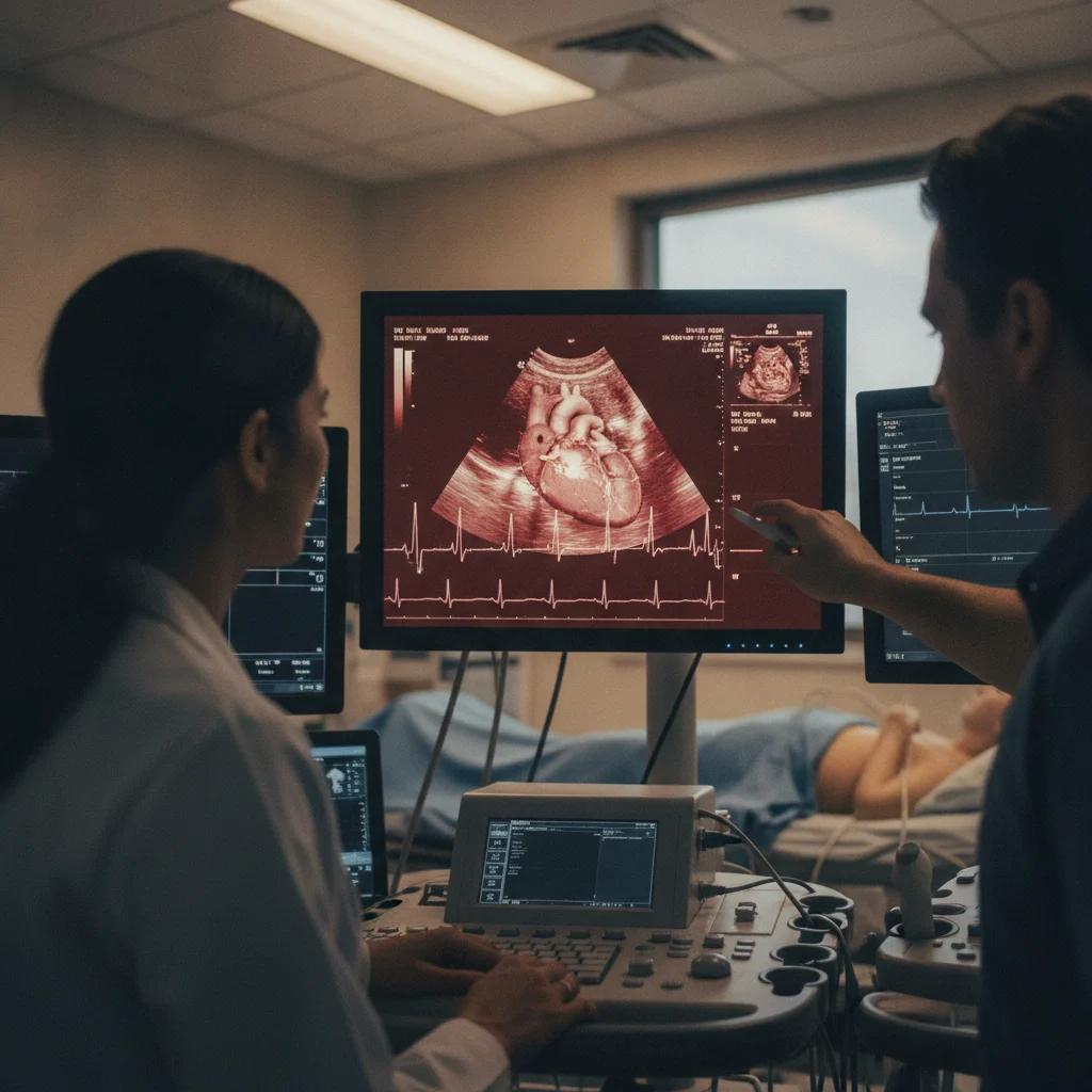
Fetal echocardiography is a specialized ultrasound technique that enables the detection of cardiac abnormalities in utero. This imaging can be performed as early as 11-14 weeks of gestation, a significant advancement compared to traditional imaging, which is typically performed at approximately 20 weeks.1
Advances in Diagnostic Modalities
Advancements in early fetal imaging have improved the identification of high-risk fetuses. This is particularly relevant for those with genetic predispositions or a family history of CHD. Earlier detection allows for earlier clinical decision-making and intervention planning. In select cases, prenatal detection facilitates in utero management of conditions such as aortic stenosis and hypoplastic left heart syndrome.1
Emerging applications of artificial intelligence (AI) have further enhanced image analysis and diagnostic accuracy in fetal echocardiography. AI tools support clinicians in the early recognition of CHD. Despite these advances, challenges persist, including variability in diagnostic accuracy during early gestation, the risk of false positives, and limited access to specialized imaging technologies. Continued integration of AI-driven tools and the development of standardized screening protocols hold promise for improving diagnostic consistency and long-term outcomes in patients with CHD.1
A separate review focused on Lipoprotein(a) in Gulf countries, navigating cardiovascular risk and therapeutic advances, did not provide specific data relevant to the diagnosis or management of aortic stenosis.2 Similarly, a study on early post-transcatheter aortic valve replacement heart failure hospitalization and outcomes in older patients did not contribute specific diagnostic or management advancements for aortic stenosis beyond the context of CHD.3
The integration of AI into fetal echocardiography primarily aims to address the inherent challenges of early gestational imaging. The small size of fetal cardiac structures and rapid heart rates during the first trimester often make manual interpretation difficult, even for experienced sonographers. AI algorithms, trained on vast datasets of both normal and abnormal fetal cardiac images, can identify subtle anomalies that might be missed by the human eye. These algorithms can automate measurements, improve image quality through noise reduction, and provide real-time diagnostic support, thereby reducing inter-observer variability and potentially shortening examination times.
Clinical Implications and Workflow Integration
The clinical implications of AI-assisted fetal echocardiography are significant. By enhancing diagnostic accuracy at earlier gestations, clinicians can provide more timely counseling to expectant parents, allowing for informed decisions regarding pregnancy management. For conditions amenable to prenatal intervention, such as critical aortic stenosis or pulmonary atresia with intact septum, earlier detection can open a wider window for therapeutic procedures, potentially improving neonatal outcomes and reducing the need for complex postnatal surgeries. Furthermore, earlier identification of severe CHD allows for planned delivery at tertiary care centers with specialized pediatric cardiology and cardiac surgery teams, optimizing immediate postnatal care.
Integrating AI tools into existing clinical workflows requires careful consideration. These systems are typically designed as decision support tools, not replacements for expert human interpretation. Sonographers and cardiologists would utilize AI outputs to augment their assessments, particularly in ambiguous cases or when screening large populations. Training healthcare professionals on the effective use of these tools, understanding their limitations, and ensuring data privacy and security are crucial for successful implementation. The goal is to create a synergistic relationship where AI enhances human capabilities, leading to more consistent and accurate diagnoses.
Limitations and Future Directions
Despite the promising advancements, several limitations warrant consideration. The performance of AI models is highly dependent on the quality and diversity of their training data. Bias in training datasets, such as an overrepresentation of certain ethnic groups or types of CHD, could lead to reduced accuracy in underrepresented populations or for rare conditions. Furthermore, the "black box" nature of some AI algorithms can make it challenging to understand the reasoning behind a particular diagnosis, which can be a barrier to clinical trust and adoption. Regulatory approval and robust validation in diverse clinical settings are essential before widespread implementation.
Future directions for AI in fetal echocardiography include the development of more sophisticated algorithms capable of dynamic analysis of cardiac function, not just structural abnormalities. Integration with other prenatal screening modalities, such as genetic testing and maternal serum markers, could create a comprehensive risk assessment platform. Longitudinal studies are needed to evaluate the long-term impact of AI-assisted early CHD detection on patient outcomes, healthcare costs, and parental psychological well-being. Continued research into explainable AI (XAI) will also be vital to foster greater transparency and clinician confidence in these advanced diagnostic tools, ultimately paving the way for a new era of precision prenatal cardiology.
The integration of artificial intelligence into fetal echocardiography marks a notable step forward in the early detection of congenital heart disease, including aortic stenosis. For clinicians, this means the potential to identify critical cardiac abnormalities significantly earlier in gestation, moving from the traditional 20-week scan to as early as 11-14 weeks. This earlier window provides a crucial opportunity for more timely counseling, intervention planning, and potentially in utero management, which could alter the disease trajectory for affected neonates. The challenge now lies in the equitable dissemination of this specialized technology and the training required to leverage AI-assisted diagnostics effectively, ensuring that these benefits are not confined to tertiary centers.
From a patient perspective, the prospect of earlier diagnosis offers both reassurance and the burden of earlier decision-making. While it allows for more comprehensive preparation and access to specialized care, it also introduces the emotional complexity of confronting a serious diagnosis earlier in pregnancy. The risk of false positives, though acknowledged, underscores the need for robust validation studies and clear communication protocols to manage parental anxiety and avoid unnecessary interventions. The industry, particularly medical device manufacturers and AI developers, must focus on creating user-friendly, reliable, and accessible platforms that can be integrated into diverse clinical settings, rather than remaining niche tools.
The current evidence, while promising, highlights the need for standardized screening protocols to ensure diagnostic consistency. Without clear guidelines for AI integration and interpretation, the variability in diagnostic accuracy could undermine the utility of these advanced tools. Future research should focus on refining AI algorithms to minimize false positives and improve accessibility, ensuring that this innovation translates into tangible improvements in long-term outcomes for all patients with CHD, not just those in well-resourced environments.
- The Pivot AI-assisted fetal echocardiography enables earlier detection of CHD, including aortic stenosis, compared to traditional imaging.
- The Data Early fetal echocardiography can detect cardiac abnormalities as early as 11-14 weeks of gestation, compared to approximately 20 weeks with traditional imaging.1
- The Action Clinicians should consider the utility of early fetal echocardiography, especially with AI integration, for high-risk pregnancies to facilitate earlier intervention planning.
ART-2026-517
06/26
Cite This Article
Team TLSFE. Ai-assisted fetal echocardiography improves early chd detection. The Life Science Feed. Updated June 24, 2026. Accessed June 24, 2026. https://thelifesciencefeed.com/cardiology/aortic-valve-stenosis/news/ai-assisted-fetal-echocardiography-improves-early-chd-detection.
Editorial & AI Standards
All content is researched from peer-reviewed, open-access sources — published trial data, clinical guidelines, and regulatory filings. AI tools are used solely to structure and summarise that evidence; no AI-generated conclusions appear without editor verification against the primary source.
Every article is reviewed by a named editor before publication. Source citations are listed in the References section. This content does not represent the views of any pharmaceutical company, medical device manufacturer, or healthcare provider.
Licence & Rights
© 2026 The Life Science Feed. All rights reserved. Unless otherwise indicated, all content is the property of The Life Science Feed and may not be reproduced, distributed, or transmitted in any form or by any means without prior written permission.
Medical Disclaimer
The information provided on The Life Science Feed is for educational and informational purposes only. It is not intended as a substitute for professional medical advice, diagnosis, or treatment. Always seek the advice of your physician or other qualified healthcare provider regarding any medical condition or treatment decision. Never disregard professional medical advice or delay in seeking it because of something you have read on this website.
References
1. Patel K, Agrawal DK. Artificial Intelligence-Assisted Fetal Echocardiography: Improving Early Detection and Neonatal Outcomes of Congenital Heart Disease. Cardiol Cardiovasc Med 2026.
2. Almahmeed W, Sabbour H, Shantouf R. Expert consensus on Lipoprotein(a) in the Gulf countries: Navigating cardiovascular risk and therapeutic advances. Atheroscler Plus 2026.
3. Kundi H, Leon M, Cohen DJ. Early Postrranscatheter Aortic Valve Replacement Heart Failure Hospitalization and Outcomes in Older Patients. J Am Heart Assoc 2026.


