Splenomegaly confronts clinicians daily, ranging from incidental findings on imaging to harbingers of serious systemic illness. Determining the etiology requires navigating a complex differential, often without clear-cut answers. A recent case report highlights isolated splenic sarcoidosis, an infrequent presentation of a disease more commonly associated with pulmonary manifestations. While rare, this case serves as a useful reminder of the breadth of possibilities and prompts a pragmatic approach to diagnosis.
This article aims to expand beyond the confines of a single case report, and provide a step-by-step approach for evaluating unexplained splenomegaly, providing clinicians with a practical framework applicable 'Monday morning'. We will focus on a systematic approach to rule out infectious, malignant, and rheumatologic causes.
Clinical Key Takeaways
lightbulb
- The PivotIsolated splenic sarcoidosis, though rare, warrants consideration in the differential diagnosis of splenomegaly, especially when other common etiologies have been excluded.
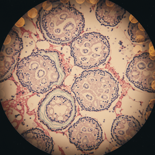
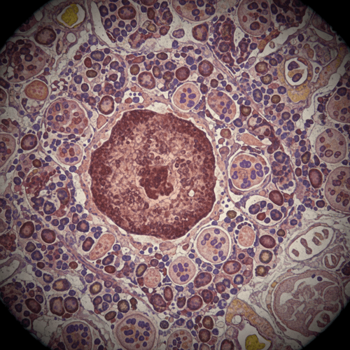
- The DataHistopathological examination of splenic tissue remains the gold standard for confirming the diagnosis, revealing non-caseating granulomas characteristic of sarcoidosis.
- The ActionImplement a structured diagnostic algorithm, beginning with excluding infectious and malignant causes, followed by considering rheumatologic and granulomatous diseases like sarcoidosis.
Diagnostic Algorithm for Splenomegaly
The initial evaluation of splenomegaly should follow a systematic approach. Begin with a thorough history and physical examination, paying close attention to potential exposures, travel history, and risk factors for infectious diseases. Laboratory investigations should include a complete blood count (CBC) with differential, liver function tests (LFTs), and a peripheral blood smear. Further testing is guided by these initial findings.
Contrast-enhanced computed tomography (CT) of the abdomen and pelvis is often the next step. This provides detailed anatomical information and can help differentiate between various causes of splenomegaly. Findings suggestive of infection include abscesses or diffuse infiltration, while masses or lymphadenopathy may point toward malignancy. In cases where malignancy is suspected, a bone marrow biopsy may be warranted.
Infectious Etiologies of Splenomegaly
Infections represent a significant proportion of splenomegaly cases. Common culprits include viral infections such as Epstein-Barr virus (EBV) and cytomegalovirus (CMV), bacterial infections like infective endocarditis and tuberculosis, and parasitic infections such as malaria and schistosomiasis. Consider these in the context of the patient's travel history and risk factors.
Testing should include serological assays for EBV and CMV, blood cultures if bacterial infection is suspected, and appropriate testing for parasitic diseases based on geographical exposure. In patients with suspected tuberculosis, consider acid-fast bacilli (AFB) staining and culture of sputum or other relevant specimens. Of course, clinicians should consider HIV testing as part of their infectious workup. This approach aligns with CDC guidelines for evaluation of fever of unknown origin and unexplained lymphadenopathy.
Malignant Etiologies of Splenomegaly
Hematologic malignancies, particularly lymphoma and leukemia, are important considerations in the differential diagnosis of splenomegaly. Lymphoma, including Hodgkin's and non-Hodgkin's lymphoma, can directly involve the spleen, leading to enlargement. Leukemias, such as chronic lymphocytic leukemia (CLL) and hairy cell leukemia, can also cause splenomegaly due to infiltration of leukemic cells.
The initial evaluation should include a careful examination for lymphadenopathy and hepatomegaly. A peripheral blood smear may reveal abnormal cells suggestive of leukemia. If lymphoma is suspected, excisional biopsy of an enlarged lymph node is the preferred diagnostic approach. The 2024 NCCN guidelines recommend PET/CT imaging for staging in many lymphoma subtypes. Bone marrow biopsy may be necessary to evaluate for leukemia or lymphoma involvement.
Rheumatologic and Other Etiologies
Sarcoidosis, an inflammatory disease characterized by the formation of granulomas in various organs, can present with isolated splenic involvement, although this is relatively rare. Other rheumatologic conditions, such as systemic lupus erythematosus (SLE) and rheumatoid arthritis, may also cause splenomegaly.
In cases of suspected sarcoidosis, a comprehensive evaluation for involvement of other organs, such as the lungs and lymph nodes, should be performed. This may include chest radiography, pulmonary function tests, and lymph node biopsy. Elevated serum angiotensin-converting enzyme (ACE) levels and soluble interleukin-2 receptor levels may support the diagnosis, but are not specific. The diagnosis of sarcoidosis requires histopathological confirmation of non-caseating granulomas in affected tissue, after excluding other causes of granulomatous disease, such as infection and malignancy. The 2020 American Thoracic Society (ATS) guidelines provide a detailed approach to diagnosing sarcoidosis.
Other less common causes of splenomegaly include storage diseases such as Gaucher disease and Niemann-Pick disease, as well as vascular disorders such as portal hypertension and splenic vein thrombosis. These conditions are typically suspected based on clinical findings and may require specialized testing for confirmation.
Caveats and Limitations
One critical limitation of relying solely on case reports is the lack of generalizability. This single instance of isolated splenic sarcoidosis may not represent the typical presentation or diagnostic course for other patients. Furthermore, the absence of a control group limits our ability to draw definitive conclusions about the efficacy of specific diagnostic strategies. The study did not discuss costs associated with extensive imaging and biopsies; this can cause significant financial toxicity for the patient.
Clinicians must be wary of confirmation bias when evaluating patients with splenomegaly. The pressure to arrive at a diagnosis can lead to premature closure and overlooking alternative explanations. A thorough and objective approach, incorporating all available clinical and laboratory data, is essential to avoid diagnostic errors.
The diagnostic algorithm outlined here requires careful coordination between primary care physicians, specialists (hematologists, infectious disease specialists, rheumatologists), and radiologists. Clear communication and efficient scheduling of tests are essential to minimize delays and avoid unnecessary procedures.
Given the extensive workup required for splenomegaly, insurance coverage and pre-authorization requirements can pose significant challenges. Clinicians should be aware of these potential barriers and advocate for their patients to ensure timely access to appropriate diagnostic testing.
Finally, it is vital to address patient anxiety and provide clear and empathetic communication throughout the diagnostic process. The uncertainty associated with splenomegaly can be distressing, and patients need reassurance and support while awaiting a definitive diagnosis.
LSF-1755318829 | December 2025

How to cite this article
Webb M. Diagnosing isolated splenic sarcoidosis a practical approach. The Life Science Feed. Published December 31, 2025. Updated December 31, 2025. Accessed March 17, 2026. https://thelifesciencefeed.com/infectious-diseases/tuberculosis/practice/diagnosing-isolated-splenic-sarcoidosis-a-practical-approach.
Copyright and license
© 2026 The Life Science Feed. All rights reserved. Unless otherwise indicated, all content is the property of The Life Science Feed and may not be reproduced, distributed, or transmitted in any form or by any means without prior written permission.
Fact-Checking & AI Transparency
This content was produced with the assistance of AI technology and has been rigorously reviewed and verified by our human editorial team to ensure accuracy and clinical relevance.
References
- Sebastiani, G., Gamberini, E., & Bombardieri, S. (2007). Splenomegaly in systemic lupus erythematosus. Lupus, 16(1), 9-13.
- James, W. E., III, & Council on Scientific Affairs (1999). The diagnosis and management of splenomegaly. JAMA, 281(12), 1126-1134.
- Costello, R. M., & Domínguez, A. R. (2023). Sarcoidosis. In StatPearls. StatPearls Publishing.