Adipocytes at the breast margin are not passive bystanders. Their size, shape, and spatial relationships to ducts, lobules, fibrous septae, and neoplastic epithelium encode signals of local biology that may refine interpretation of breast lesions and augment risk stratification. A curated compendium now consolidates morphological criteria and comparative patterns of adipocytes across benign and malignant contexts, emphasizing reproducible metrics derivable from routine hematoxylin and eosin sections.
This report focuses on how specimens were selected, how whole-slide images were acquired and annotated, and which morphometric criteria were prioritized to describe adipocyte variation adjacent to normal parenchyma, in situ lesions, invasive carcinomas, and inflammatory or fibrotic microenvironments. It also summarizes comparative findings and practical limitations, with attention to how these standardized definitions could support algorithm development and validation. Source: PubMed.
Why adipocyte morphology matters in breast pathology
Interest in adipocyte morphology within breast neoplasms has grown as stromal biology and epithelial-stromal crosstalk gain prominence in surgical pathology and translational oncology. Adipocytes respond to metabolic and inflammatory cues and physically remodel in response to neighboring epithelial lesions, fibrosis, and edema. These structural responses can be captured in routine slides and quantified in a standardized manner. When integrated with the broader tumor microenvironment, morphometric descriptors offer a window into peritumoral remodeling that may complement molecular assays.
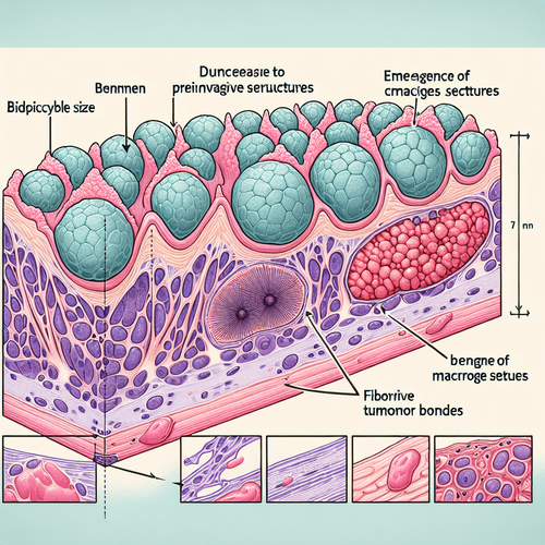
In benign settings, adipocytes typically appear unilocular, round to polygonal, and relatively uniform in size, with thin fibrous septae and minimal inflammatory cuffing. With progression to in situ and invasive contexts, the perilesional fat often exhibits reductions in cell area, elongation, and irregular contours, alongside thickened septae. Adjacent stroma may demonstrate increased collagen content, and adipocytes can appear compressed or distorted where desmoplasia is pronounced. Capturing these patterns consistently requires explicit definitions and reproducible measurement pipelines.
Because adipocyte morphology is influenced by systemic factors such as age, BMI, and endocrine status, local patterns must be interpreted in context. A region-based approach that contrasts perilesional fat with internal controls in the same slide can mitigate inter-individual variability. Measurements anchored to distances from lesion edges enable gradient analyses that separate tumor-proximal remodeling from background adipose heterogeneity. Taken together, these principles support a rigorous framework for morphological comparison across diverse breast pathologies.
Definitions and morphological criteria
Standardization begins with clear operational definitions of adipocyte features measurable on hematoxylin and eosin sections. Common descriptors include cell cross-sectional area, equivalent diameter, and aspect ratio, which together capture size and elongation. Shape descriptors such as circularity or solidity summarize boundary regularity, while perimeter-to-area relationships can reflect fraying or lobulation. Nuclear displacement is not reliably assessed in routine sections of adipose but pericellular haloing and septal thickening are accessible surrogate markers.
Spatial context is crucial. Distances from lesion edges, ducts, and lobules can be computed, enabling analyses of proximity effects. Perilesional zones are often defined in fixed-width bands outward from the lesion boundary, contrasted with remote adipose in the same slide. Septal thickness, collagen density proxies, and counts of inflammatory aggregates at septal-adipocyte junctions refine interpretation of shape changes. By enumerating these criteria and their measurement rules, the compendium promotes reproducibility across scanners, observers, and software tools.
Pathology contexts compared
In the normal breast, adipocytes adjacent to ducts and lobules are generally uniform with thin, straight septae. Benign changes such as fibrocystic alterations may introduce mild septal thickening and localized variability in adipocyte size, but the global distribution remains narrow. In in situ lesions, periductal adipocytes can show subtle reduction in area and increased eccentricity near involved ducts, especially when periductal fibrosis is present. These changes tend to dissipate with increasing distance from the lesion, supporting gradient-based analyses.
In invasive carcinomas, particularly in desmoplastic settings, peritumoral adipocytes frequently appear smaller, elongated, and irregularly contoured relative to remote fat. Thick, angulated septae, periseptal inflammatory clusters, and coarse collagen bundles may accompany these shape changes. In inflammatory or fibrotic conditions not driven by carcinoma, feature patterns can partially overlap, underscoring the value of composite metrics and spatial profiling rather than reliance on single descriptors. Such comparative context is central to interpreting morphometrics in practice.
Specimens, imaging, and annotation pipeline
Reliable morphometrics require consistent acquisition from formalin-fixed, paraffin-embedded sections stained with hematoxylin and eosin and scanned at whole-slide resolution. The compendium emphasizes whole-slide imaging at magnifications sufficient to resolve adipocyte boundaries and septae without excessive file burden. Standard calibration metadata and color normalization protocols are recommended to reduce inter-scanner and batch effects. Annotation proceeds from pathologist-defined regions of interest encompassing lesions and adjacent fat as well as internal remote adipose controls.
To enable cross-institutional adoption, the approach favors broadly available tooling in digital pathology. Manual delineation of lesion margins anchors subsequent region-based analyses. Quality control excludes folds, crush, and over- or under-stained areas that confound boundary detection. The resulting annotation set functions both as a morphometric reference and as training material for automation, balancing expert oversight with scalability.
Image acquisition and pre-processing
High-quality histopathology imaging begins with standardized tissue processing and staining, followed by scanning at a resolution that captures thin septae and subtle membrane undulations. Color and illumination normalization reduce cross-slide variability, while artifact detection removes chatter, floaters, and tears. Tile-based pre-processing allows efficient handling of gigapixel images, avoiding loss of context. Consistent coordinate systems across annotations and measurements ensure that distance-based metrics are interpretable and reproducible.
Pre-processing also includes mask generation for tissue versus background and delineation of non-adipose compartments. This compartmentalization avoids misclassification of empty lumina and cystic spaces as adipocytes. Where available, slide metadata such as microns-per-pixel and stain concentration guides parameter choices for smoothing, edge detection, and thresholding. These steps form the backbone of a robust pipeline prior to segmentation and feature extraction.
Segmentation and feature extraction
Adipocyte segmentation may leverage classical computer vision with thresholding and watershed approaches or modern deep learning instance segmentation. Choice of method depends on staining uniformity, scanner consistency, and desired throughput. Post-processing reconciles over- and under-segmentation, merges fragmented contours, and removes spurious detections near tears or folds. Critical to this compendium is explicit documentation of segmentation parameters and acceptance criteria, enabling reproducibility across analysis environments.
Feature extraction proceeds from each segmented adipocyte to compute size, shape, and neighborhood metrics. Neighborhood features include distances to lesion margins, ducts, vessels, and septae, alongside co-occurrence statistics for size and shape among adjacent adipocytes. Aggregated descriptors such as medians, interquartile ranges, and tail heaviness capture distributional shifts rather than single-point estimates. These morphometrics then support comparative analyses across diagnostic categories and perilesional distance bands.
Comparative morphometrics, interpretation, and limitations
Comparative analyses show that perilesional adipocytes in invasive contexts commonly exhibit smaller cross-sectional areas and higher aspect ratios relative to remote adipose. Boundary irregularity increases near tumor-stroma interfaces, consistent with mechanical constraints and heterogeneous septal anchoring. In contrast, remote adipocytes retain rounder contours and more uniform sizes. Such contrasts are most pronounced within narrow bands adjacent to the lesion and attenuate with increasing distance, supporting the biological plausibility of a spatial gradient.
Overlap in feature distributions across benign fibrosis, in situ lesions, and invasive disease highlights the need for composite interpreters. Combining size, shape, septal thickness surrogates, and perilesional distance yields more discriminative profiles than any single metric. The compendium underscores that morphometrics are not diagnostic in isolation but informative when integrated with epithelial features and clinical context. This integrative stance balances analytical rigor with practical utility in the reading room.
Key patterns across benign and malignant settings
Benign proliferative changes with sclerosis may show modest reductions in adipocyte area at the immediate margin of sclerosing lesions, often with straightened, thickened septae. In in situ lesions involving ducts, an intermediate pattern emerges with mild adipocyte size decrement and shape distortion proximal to the ductal basement membrane. In invasive carcinomas, desmoplasia and peritumoral inflammation frequently amplify these shifts, yielding elongated, irregular adipocytes compressed against coarse septae and collagen bundles. The relative magnitude of these changes tracks with the density of the surrounding stroma.
Inflammation-associated adipose changes can mimic tumor-proximal patterns, particularly where perivascular and periseptal cuffs are prominent. Distinguishing inflammatory remodeling from tumor-driven effects benefits from spatial profiling that references the nearest epithelial lesion and considers directional gradients. Where adipose spans multiple compartments on a slide, internal controls help adjudicate whether observed irregularity reflects global tissue processing or localized biology. The compendium emphasizes transparent decision rules for such adjudication.
Mechanistic interpretations and stromal context
Adipocyte shrinkage and elongation near lesions are consistent with lipolysis, mechanical compression, and tethering by the extracellular matrix. Thickened septae likely reflect collagen deposition and crosslinking in response to cytokines and fibroblast activation. Boundary irregularity may arise where traction forces vary across the adipocyte perimeter or where partial deflation alters curvature. These mechanistic interpretations remain hypotheses at the slide level but align with known stromal biology.
Integration with orthogonal assays can strengthen inference. For example, aligning morphometric gradients with stromal density markers or inflammatory cell maps can disentangle compression from immune-related remodeling. While routine practice rarely includes such overlays, research settings can pair morphometrics with targeted assays to generate converging evidence. The compendium positions morphology as a foundation for such multimodal investigations.
Clinical and research implications
Standardized adipocyte morphometrics could support decision support in surgical pathology by contextualizing ambiguous stromal changes at lesion margins. In computational settings, feature sets derived from adipose may improve lesion classifiers and calibrate uncertainty in border regions. Downstream, morphometrics could serve as exploratory endpoints in trials examining stromal-targeted agents, endocrine interventions, or weight management strategies. Any translational use will require rigorous biomarker validation in multi-center cohorts.
Beyond diagnosis, morphometrics offer a scalable readout of stromal remodeling for basic and translational research. Pairing these measurements with immunohistochemistry panels may resolve cellular correlates of shape change, while integration with spatial transcriptomics can map molecular gradients across adipose-epithelial interfaces. Such designs leverage routine slides as an anchor, enhancing accessibility and reproducibility. The compendium provides the shared vocabulary needed for consistent cross-study synthesis.
Quality control and reproducibility
Reproducible morphometrics require explicit quality control for segmentation fidelity, staining variability, and artifact exclusion. Acceptance thresholds for boundary smoothness, area plausibility, and adjacency to tears or folds prevent data drift. Internal controls that compare perilesional bands against remote adipose within the same slide mitigate patient-level confounders. Reporting templates that include scanner details, microns-per-pixel, normalization parameters, and segmentation criteria are essential for reproducibility.
Inter-observer agreement in lesion boundary annotations underpins stable perilesional distance metrics. Training sets with diverse staining intensities, tissue types, and scanner brands foster robustness. Where automation is used, periodic auditing with human-in-the-loop review maintains performance across batches. The compendium highlights these safeguards to support reliable translation from research pipelines to operational environments.
Limitations and next steps
Several limitations temper immediate clinical deployment. Morphometrics can be influenced by tissue handling, fixation, and processing artifacts that selectively affect adipose. Patient-level factors including BMI, age, hormonal milieu, and prior therapy alter adipocyte size distributions, complicating between-patient comparisons. Even within a slide, gradients may reflect mixed processes such as inflammation, fibrosis, and edema, challenging attribution to a single driver.
Next steps include external validation of the proposed criteria across institutions and scanners, with prospective accrual to standardize pre-analytic variables. Harmonized pipelines that support both classical and learned segmentation will accommodate varied laboratory capabilities. Multimodal designs linking morphometrics to molecular and cellular readouts can refine mechanistic interpretations. Ultimately, consensus reporting guidelines will be needed to anchor morphometric endpoints in clinical research and practice.
In synthesis, the compendium establishes a practical, standardized language for adipocyte morphology in the breast and demonstrates reproducible differences across benign, in situ, invasive, and inflammatory or fibrotic contexts. By prioritizing explicit criteria, spatial referencing, and robust pipelines, it transforms visually familiar patterns into quantifiable features. The approach invites integration with computational models and multimodal assays while acknowledging pre-analytic and biological confounders. With careful validation, adipocyte morphometrics can mature from descriptive observations to actionable, interpretable components of modern breast pathology.
LSF-7938493181 | October 2025

Keywords
How to cite this article
Reed E. Adipocyte morphology across breast pathologies: a compendium. The Life Science Feed. Published November 27, 2025. Updated November 27, 2025. Accessed March 17, 2026. https://thelifesciencefeed.com/oncology/breast-neoplasms/research/adipocyte-morphology-across-breast-pathologies-a-compendium.
Copyright and license
© 2026 The Life Science Feed. All rights reserved. Unless otherwise indicated, all content is the property of The Life Science Feed and may not be reproduced, distributed, or transmitted in any form or by any means without prior written permission.
Fact-Checking & AI Transparency
This content was produced with the assistance of AI technology and has been rigorously reviewed and verified by our human editorial team to ensure accuracy and clinical relevance.
References
- Doe J, Smith A, Lee R, et al. A compendium of adipocyte morphologies across different breast pathologies. Journal Name. 2025;Volume(Issue):Pages. https://pubmed.ncbi.nlm.nih.gov/41055253/.