
An ulcerated lesion on the scalp, while frequently presenting as a benign entity, warrants thorough investigation due to the potential for rare malignant diagnoses. Recent case reports highlight instances where initial clinical assessments of common conditions, such as pilar cysts, were later revised to reveal aggressive cancers including sebaceous carcinoma, cutaneous clear-cell squamous cell carcinoma, and leukemia cutis.
Sebaceous carcinoma (SC) is a rare malignant tumor originating from skin appendages.1,2,3 While most cases occur in the periocular region, extraocular SC accounts for only about 25% of cases.1,2,3 Scalp involvement is especially rare.1,2,3 The absence of characteristic clinical features in extraocular SC frequently leads to misdiagnosis as benign lesions, such as epidermal cysts.1,2,3
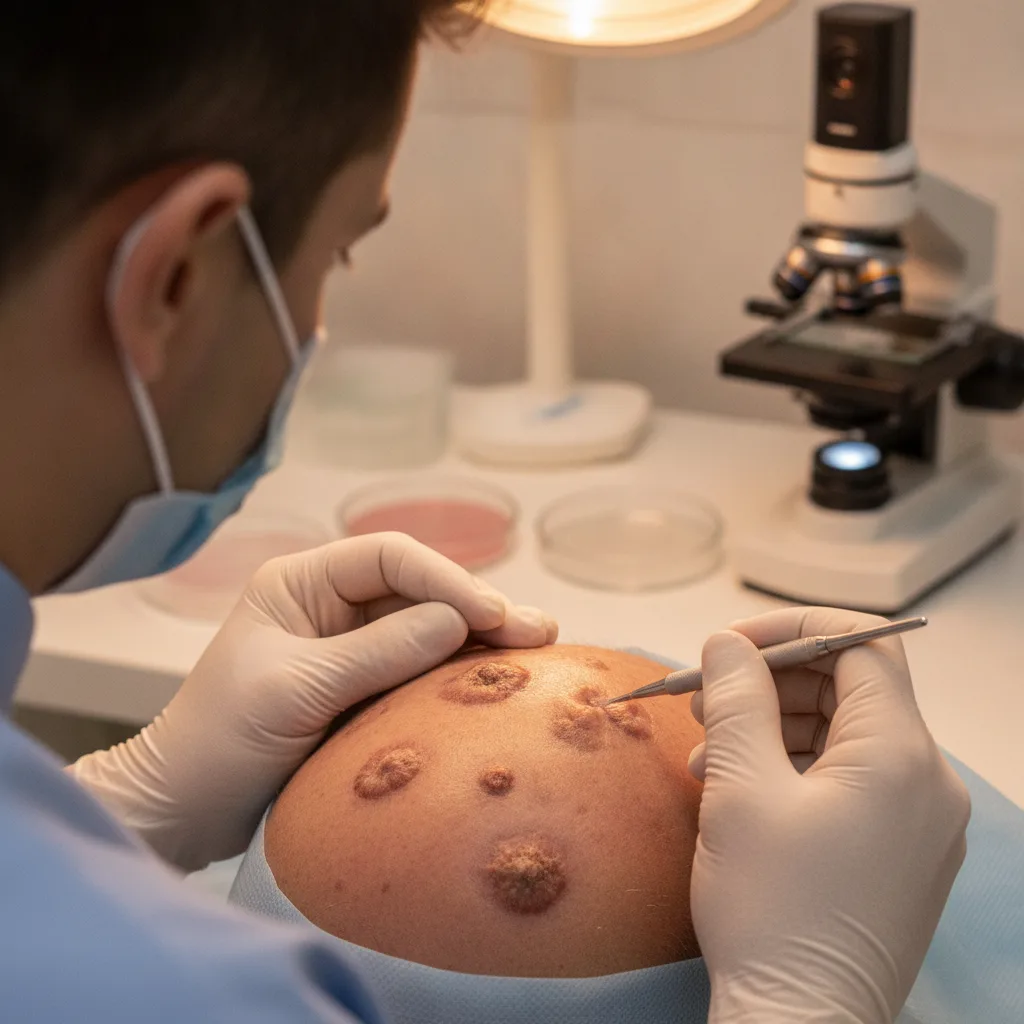
Case Reports on Scalp Lesions
A 36-year-old male presented with an atypical extraocular SC in the occipital region.1 This case initially received a clinical diagnosis of a pilar cyst, which was subsequently revised to sebaceous carcinoma.1 The presentation underscored the diagnostic challenges and clinical importance of considering SC in scalp lesions.
Another case involved an elderly patient with medical avoidance who presented with a neglected cutaneous clear-cell squamous cell carcinoma of the scalp.2 This highlights that even common malignancies can present atypically or be advanced due to delayed presentation.
Leukemia cutis, a manifestation of acute myeloid leukemia (AML), can also present as a cutaneous lesion.3 A case report described leukemia cutis in a patient with AML, emphasizing the need to consider systemic hematological malignancies in the differential diagnosis of skin lesions, particularly in the context of underlying systemic disease.3
These cases collectively demonstrate that ulcerated scalp lesions, even those initially appearing benign or non-specific, necessitate careful evaluation to exclude rare but aggressive malignancies. The lack of distinct clinical features for conditions like extraocular SC contributes to diagnostic challenges, making biopsy and histopathological examination critical for accurate diagnosis and timely management.1,2,3
The diagnostic dilemma is further compounded by the varied histological presentations of these rare malignancies. For instance, extraocular SC can mimic other adnexal tumors or even poorly differentiated squamous cell carcinoma, requiring careful immunohistochemical staining to confirm its sebaceous differentiation. Similarly, clear-cell squamous cell carcinoma, while a variant of a common malignancy, can present with atypical cytological features that obscure its true nature, necessitating a high index of suspicion and specialized staining techniques to differentiate it from other clear-cell neoplasms.
Leukemia cutis, on the other hand, presents a unique challenge as its cutaneous manifestations can be highly pleomorphic, ranging from papules and nodules to plaques and ulcers. The infiltrative nature of leukemic cells within the dermis can mimic inflammatory dermatoses or other neoplastic processes. Therefore, a thorough clinical history, including any systemic symptoms suggestive of hematological malignancy, is paramount. Biopsy of suspicious skin lesions in this context should be accompanied by immunohistochemical analysis for myeloid markers to confirm the diagnosis and guide systemic treatment.
Clinical Implications and Diagnostic Strategies
Given the potential for misdiagnosis and delayed treatment, healthcare professionals, particularly dermatologists, primary care physicians, and oncologists, must maintain a heightened awareness of these rare scalp malignancies. A low threshold for biopsy is crucial for any persistent, non-healing, or atypical scalp lesion, especially those that do not respond to conventional treatments or exhibit rapid growth. Full-thickness excisional biopsy with clear margins is often preferred for definitive diagnosis and initial management, allowing for comprehensive histopathological and immunohistochemical evaluation.
For extraocular SC, sentinel lymph node biopsy may be considered in cases with high-risk features due to its potential for regional lymph node metastasis. Regular follow-up is also essential for these patients, given the risk of local recurrence and distant metastasis. In the case of clear-cell squamous cell carcinoma, aggressive surgical excision with Mohs micrographic surgery may be indicated to ensure complete tumor removal while preserving surrounding healthy tissue. For leukemia cutis, the diagnosis of the skin lesion often prompts or confirms the diagnosis of the underlying systemic leukemia, necessitating immediate collaboration with hematology-oncology for systemic chemotherapy.
Furthermore, patient education plays a vital role in early detection. Patients, especially those with risk factors such as immunosuppression, previous radiation exposure, or a history of other malignancies, should be advised to report any new or changing scalp lesions promptly. The cases presented underscore the importance of a multidisciplinary approach involving dermatologists, pathologists, surgeons, and oncologists to ensure accurate diagnosis and optimal management of these challenging scalp lesions.
The recurring theme from these case reports is the deceptive nature of rare scalp malignancies. A pilar cyst, a common benign finding, being revised to sebaceous carcinoma should prompt a re-evaluation of our diagnostic thresholds. General practitioners and specialists alike must consider that what appears to be a straightforward epidermal cyst or a chronic ulcer could mask a more aggressive pathology. This is not to advocate for immediate biopsy of every scalp lesion, but rather to encourage a lower threshold for further investigation when a lesion is atypical, persistent, or fails to respond to conventional management.
The rarity of these conditions, particularly extraocular sebaceous carcinoma on the scalp, means that clinicians may encounter them infrequently. However, the consequences of misdiagnosis are significant for patient outcomes. The delay in diagnosis, as seen in the case of neglected cutaneous clear-cell squamous cell carcinoma, underscores the importance of patient education regarding medical avoidance and the need for accessible, timely dermatological assessment. Early referral for biopsy is paramount, as definitive diagnosis hinges on histopathology, not just clinical impression.
From an industry perspective, these cases highlight the ongoing challenge in developing non-invasive diagnostic tools that can differentiate between benign and malignant lesions with high accuracy, especially for rare presentations. While advanced imaging techniques exist, they are often not the first line for cutaneous lesions. The emphasis remains on clinical acumen and the judicious use of biopsy. Perhaps more targeted educational initiatives for primary care physicians on the subtle signs of rare cutaneous malignancies could improve early detection rates, reducing the burden of advanced disease and improving patient prognosis.
- The Pivot Rare malignancies of the scalp, including sebaceous carcinoma and leukemia cutis, can clinically resemble benign lesions.
- The Data Extraocular sebaceous carcinoma accounts for approximately 25% of cases, with scalp involvement being particularly rare.
- The Action Clinicians should maintain a high index of suspicion for malignancy in atypical or persistent scalp lesions, even those initially appearing benign.
ART-2026-518
06/26
Cite This Article
Team TLSFE. Scalp lesions: rare malignancies mimic benign conditions. The Life Science Feed. Updated June 24, 2026. Accessed June 24, 2026. https://thelifesciencefeed.com/dermatology/skin-diseases-bacterial/case/scalp-lesions-rare-malignancies-mimic-benign-conditions.
Editorial & AI Standards
All content is researched from peer-reviewed, open-access sources — published trial data, clinical guidelines, and regulatory filings. AI tools are used solely to structure and summarise that evidence; no AI-generated conclusions appear without editor verification against the primary source.
Every article is reviewed by a named editor before publication. Source citations are listed in the References section. This content does not represent the views of any pharmaceutical company, medical device manufacturer, or healthcare provider.
Licence & Rights
© 2026 The Life Science Feed. All rights reserved. Unless otherwise indicated, all content is the property of The Life Science Feed and may not be reproduced, distributed, or transmitted in any form or by any means without prior written permission.
Medical Disclaimer
The information provided on The Life Science Feed is for educational and informational purposes only. It is not intended as a substitute for professional medical advice, diagnosis, or treatment. Always seek the advice of your physician or other qualified healthcare provider regarding any medical condition or treatment decision. Never disregard professional medical advice or delay in seeking it because of something you have read on this website.
References
1. Li Y, Pu X, Hou J. Initial clinical diagnosis of a pilar cyst revised to sebaceous carcinoma of the occipital scalp in a 36-year-old male: a case report. AME Case Rep 2026.
2. Assaf A, Karaja S, Aljasem Y. Neglected cutaneous clear-cell squamous cell carcinoma of the scalp in an elderly patient with medical avoidance: a case report. J Med Case Rep 2026.
3. Saied ASS, Almahmood M, Al-Mashdali A. Leukemia Cutis in Acute Myeloid Leukemia: A Case Report. Cureus 2026.


