The quest for efficient osteoporosis screening methods continues, especially for patients who may not be flagged through traditional risk assessment. A recent multi-center study explored the potential of using cranial computed tomography (CT) scans, routinely performed for other indications, to predict major osteoporotic fractures based on Hounsfield unit (HU) measurements. The appeal is obvious: no additional radiation exposure, no extra cost for the scan itself. But does this approach hold water, and more importantly, how does it fit into existing clinical workflows?
This letter to the editor addresses some valid questions and concerns raised about the original study, focusing on methodology and patient selection. While the authors present a compelling case for opportunistic screening, we need to examine the practical implications and potential pitfalls of widespread implementation.
Clinical Key Takeaways
lightbulb
- The PivotCranial CT scans, typically used for neurological assessments, may offer an opportunity for opportunistic osteoporosis screening by measuring Hounsfield Units.
- The DataThe original study demonstrated that CT-derived HU measurements could predict major osteoporotic fractures, suggesting a potential for identifying at-risk individuals.
- The ActionExplore the feasibility of integrating HU measurements into routine CT reporting workflows, but be mindful of patient selection biases and the need for further validation studies.
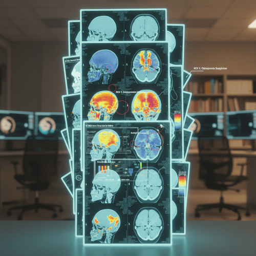
The Promise of Opportunistic Screening
Opportunistic screening, the practice of using existing data or tests to identify unrelated health risks, is gaining traction in various fields. In this case, the idea is to leverage cranial CT scans, primarily ordered for neurological assessments, to simultaneously assess bone density and predict fracture risk. The original study, which this letter responds to, found a correlation between HU measurements derived from CT scans and the likelihood of major osteoporotic fractures. This suggests that a simple, automated analysis of existing images could flag patients who warrant further investigation and potential intervention.
Guideline Context: Where Does This Fit?
Current guidelines, such as those from the National Osteoporosis Foundation (NOF), recommend bone density testing (DXA scan) for women aged 65 and older, and for younger women and men with risk factors. This opportunistic CT screening does *not* replace DXA. Instead, it could act as a pre-screen, identifying individuals who might benefit from a formal DXA scan. If a patient is already undergoing a CT for other reasons, extracting HU data presents a low-risk, low-cost method to potentially improve risk stratification. The key is to remember that this is a screening tool, not a diagnostic replacement. Further research is needed to define clear thresholds and algorithms for referral to DXA based on CT-derived HU values. It must be emphasized that, as of today, there is no official endorsement of this method within accepted clinical practice guidelines for osteoporosis.
Methodological Concerns
While the concept is appealing, several methodological concerns need addressing. The patient population undergoing cranial CT scans is inherently different from the general population. These individuals often present with neurological symptoms, head trauma, or other conditions that might independently influence bone density. This selection bias could skew the results and limit the generalizability of the findings. The letter to the editor rightly points out the need for careful consideration of patient demographics and comorbidities when interpreting HU measurements. Furthermore, variations in CT scanner technology, imaging protocols, and measurement techniques could introduce variability and affect the reproducibility of results. The authors acknowledge these limitations and emphasize the need for standardized protocols and further validation studies across diverse populations.
The AI Angle
The true potential of this approach lies in the application of artificial intelligence (AI). Automating HU measurements and integrating alerts into Picture Archiving and Communication Systems (PACS) or Electronic Health Record (EHR) systems could streamline the screening process and minimize the burden on radiologists. Imagine an AI algorithm that automatically analyzes cranial CT scans, calculates HU values, and flags patients at high risk of osteoporosis for further evaluation. This could significantly improve the efficiency of osteoporosis screening and facilitate earlier intervention. However, the development and validation of such AI algorithms require large, well-curated datasets and rigorous testing to ensure accuracy and reliability. The cost of deploying and maintaining these AI systems also needs consideration.
Economic Considerations
The economic argument for opportunistic osteoporosis screening is compelling. By leveraging existing CT scans, we can potentially identify at-risk individuals without incurring the cost of additional imaging. However, the downstream costs associated with further evaluation, such as DXA scans, specialist consultations, and treatment, need to be factored in. Moreover, the implementation of this screening program requires investment in infrastructure, including AI software, data storage, and staff training. The potential for false positives and unnecessary investigations also needs to be carefully weighed against the benefits of early detection. A thorough cost-effectiveness analysis is essential to determine the overall value of this approach. Furthermore, it remains unclear whether insurance providers will reimburse for the additional analysis of CT scans for opportunistic osteoporosis screening. The lack of dedicated billing codes may pose a barrier to widespread adoption.
The clinical implications of opportunistic osteoporosis screening using cranial CT scans are multifaceted. If validated and implemented effectively, it could lead to earlier detection and treatment of osteoporosis, potentially reducing the incidence of major osteoporotic fractures. However, clinicians need to be aware of the limitations of this approach and avoid over-interpreting HU measurements. Integrating this screening program into existing clinical workflows requires careful planning and coordination between radiologists, primary care physicians, and other healthcare professionals. Consider the potential impact on workflow. Will radiology departments bear the brunt of additional data analysis? Will primary care physicians be prepared to manage the influx of patients flagged by this screening method? These logistical considerations are paramount. The liability implications of *not* acting on these incidental findings also need examination. It must be documented clearly that the patient has been notified and referred appropriately when indicated.
LSF-8985290167 | January 2026

How to cite this article
O'Malley L. Can routine ct scans predict osteoporosis risk?. The Life Science Feed. Published January 25, 2026. Updated January 25, 2026. Accessed April 2, 2026. https://thelifesciencefeed.com/endocrinology/osteoporosis/innovation/can-routine-ct-scans-predict-osteoporosis-risk.
Copyright and license
© 2026 The Life Science Feed. All rights reserved. Unless otherwise indicated, all content is the property of The Life Science Feed and may not be reproduced, distributed, or transmitted in any form or by any means without prior written permission.
Fact-Checking & AI Transparency
This content was produced with the assistance of AI technology and has been rigorously reviewed and verified by our human editorial team to ensure accuracy and clinical relevance.
References
- Kanis, J. A., et al. "European guidance for the diagnosis and management of osteoporosis in postmenopausal women." Osteoporosis International 24.1 (2013): 23-57.
- Cosman, F., et al. "Clinician's Guide to Prevention and Treatment of Osteoporosis." Osteoporosis International 25.10 (2014): 2359-2381.
- Siris, E. S., et al. "Identification and fracture outcomes of undiagnosed low bone mineral density in postmenopausal women: results from the National Osteoporosis Risk Assessment (NORA)." JAMA 290.12 (2003): 1575-1583.
- Popp, A. W., et al. "Accuracy of opportunistic osteoporosis screening on routine CT scans of the chest and abdomen." European Radiology 28.11 (2018): 4745-4752.