
Background
The push for osteoporosis screening is intensifying, driven by the desire to prevent debilitating fractures. Current guidelines, such as those from the National Osteoporosis Foundation (NOF), recommend bone mineral density (BMD) testing using dual-energy X-ray absorptiometry (DXA) for women aged 65 and older and men aged 70 and older, as well as for younger individuals with specific risk factors. However, DXA scans require dedicated equipment and personnel, creating logistical hurdles, especially in resource-limited settings.
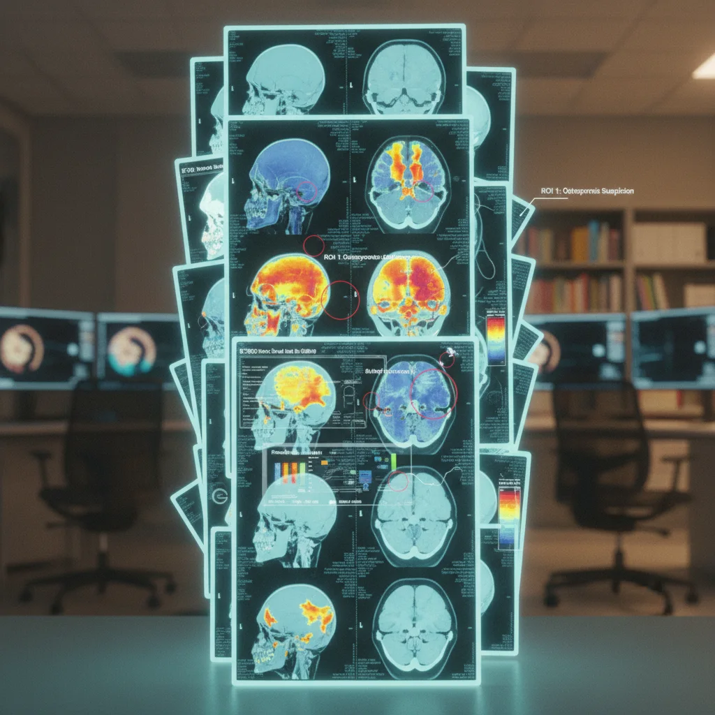
The idea of leveraging existing CT scans, often performed for other clinical indications, for opportunistic osteoporosis screening is thus attractive. Cranial CT scans, readily available in many emergency departments and radiology suites, could potentially provide valuable information about bone density. The Hounsfield unit (HU), a quantitative measure of radiodensity, can be extracted from these scans and correlated with BMD. The original study explored this concept across multiple centers, aiming to establish a reliable threshold for predicting major osteoporotic fractures.
This approach, if validated, could significantly expand screening coverage, particularly among individuals who might not otherwise undergo DXA testing. However, the devil is always in the details, particularly concerning methodological rigor and potential biases.
Authors' Response
The authors of the original study addressed several key criticisms in their response. A primary concern raised by the letter to the editor was the potential for confounding variables to influence the relationship between cranial CT-derived HU and fracture risk. Specifically, the critics questioned whether factors such as age, sex, and comorbidities were adequately controlled for in the analysis.
The authors countered by stating that they had indeed adjusted for these variables in their statistical models. However, the extent and nature of these adjustments remain somewhat unclear. For instance, while they mention adjusting for age, it's not specified whether this was done using linear or non-linear terms, which could significantly impact the results, especially given the complex relationship between age and bone density.
Another point of contention was the generalizability of the findings. The study population may not be representative of the broader population at risk for osteoporosis. The authors acknowledged this limitation to some degree, emphasizing the need for further research in diverse populations.
The response also touched upon the issue of reproducibility. The critics questioned whether the HU measurements obtained from different CT scanners and protocols were comparable. The authors defended their methodology by highlighting the use of standardized protocols and quality control measures across participating centers. Yet, inter-scanner variability remains a potential source of bias that warrants further investigation.
Limitations
Despite the authors' attempts to address the criticisms, several limitations remain. First, the retrospective design of the study introduces the possibility of selection bias. Patients undergoing cranial CT scans may differ systematically from those who do not, potentially skewing the results. A prospective study design would be more robust, but also more resource-intensive.
Second, the sample size, while multi-center, may still be insufficient to detect subtle but clinically significant associations. Furthermore, the reliance on cranial CT scans, rather than dedicated BMD measurements, introduces inherent inaccuracies. Cranial bone density may not perfectly reflect bone density at other skeletal sites, such as the hip or spine, which are more commonly associated with osteoporotic fractures.
Third, the study's reliance on Hounsfield units as a surrogate marker for BMD raises concerns about measurement error and calibration issues. While the authors mention using standardized protocols, the potential for inter-scanner variability cannot be completely eliminated. Furthermore, the relationship between HU and BMD may vary depending on the specific CT scanner and reconstruction algorithm used.
Finally, and perhaps most importantly, the study does not address the cost-effectiveness of opportunistic osteoporosis screening using cranial CT scans. While the approach may be convenient and readily accessible, the added cost of analyzing CT images for bone density, as well as the potential for false-positive results leading to unnecessary further testing, must be carefully considered. Who, ultimately, will pay for this? The current DRG reimbursement models do not account for it.
Clinical Implications
The allure of using existing cranial CT scans for osteoporosis screening lies in its potential to reach a broader population at risk. However, before this approach can be widely adopted, several practical considerations must be addressed.
First, clear protocols and guidelines are needed to ensure standardized HU measurements and interpretation across different radiology departments. This would require training radiologists and technologists in the proper techniques for extracting and analyzing bone density data from CT images.
Second, appropriate referral pathways must be established to ensure that patients identified as being at high risk for osteoporosis are promptly referred for further evaluation and treatment. This would involve coordinating care between radiologists, primary care physicians, and specialists such as endocrinologists or rheumatologists.
Third, the economic implications of implementing opportunistic osteoporosis screening using cranial CT scans must be carefully evaluated. This would require assessing the cost of analyzing CT images, the potential savings from preventing fractures, and the impact on healthcare resource utilization.
Widespread adoption hinges on addressing these workflow and financial concerns. Without clear reimbursement models and standardized protocols, this approach, despite its potential, risks becoming another unfunded mandate that strains already overburdened healthcare systems.
lightbulb
- The PivotOpportunistic osteoporosis screening via cranial CT Hounsfield Units is promising, but requires further validation before widespread implementation.
- The DataThe original study demonstrated a significant association between cranial CT-derived HU and major osteoporotic fractures, but the letter raised concerns about potential confounding factors.
- The ActionClinicians should remain cautious when using cranial CT HU values for osteoporosis screening until larger, prospective studies confirm its reliability and accuracy.
ART-2026-51
06/26
Cite This Article
Team E. Opportunistic osteoporosis screening from ct scans a validated approach?. The Life Science Feed. Published January 1, 2026. Updated June 28, 2026. Accessed July 18, 2026. https://thelifesciencefeed.com/orthopedics/osteoporosis/research/opportunistic-osteoporosis-screening-from-ct-scans-a-validated-approach.
Editorial & AI Standards
All content is researched from peer-reviewed, open-access sources: published trial data, clinical guidelines, and regulatory filings. AI tools are used solely to structure and summarise that evidence; no AI-generated conclusions appear without editor verification against the primary source.
Every article is reviewed by a named editor before publication. Source citations are listed in the References section. This content does not represent the views of any pharmaceutical company, medical device manufacturer, or healthcare provider.
Licence & Rights
© 2026 The Life Science Feed. All rights reserved. Unless otherwise indicated, all content is the property of The Life Science Feed and may not be reproduced, distributed, or transmitted in any form or by any means without prior written permission.
Medical Disclaimer
The information provided on The Life Science Feed is for educational and informational purposes only. It is not intended as a substitute for professional medical advice, diagnosis, or treatment. Always seek the advice of your physician or other qualified healthcare provider regarding any medical condition or treatment decision. Never disregard professional medical advice or delay in seeking it because of something you have read on this website.
References
- Kanis, J. A., et al. "European guidance for the diagnosis and management of osteoporosis in postmenopausal women." Osteoporosis International 30.1 (2019): 3-44.
- Cosman, F., et al. "Clinician’s Guide to Prevention and Treatment of Osteoporosis." Osteoporosis International 25.10 (2014): 2359-2381.
- Lewiecki, E. M., et al. "2023 Clinician’s Guide to Prevention and Treatment of Osteoporosis." Journal of Clinical Densitometry 26.3 (2023): 101304.


