
The increasing complexity of pulmonary diagnostics and interventions necessitates proficient bronchoscopic skills among pulmonologists and thoracic surgeons. The American Thoracic Society (ATS) 2026 conference will address this by offering a comprehensive, hands-on guide to basic bronchoscopy, endobronchial ultrasound (EBUS), and guided bronchoscopy techniques.
Bronchoscopy remains a cornerstone of pulmonary medicine, facilitating direct visualization of the tracheobronchial tree, tissue sampling, and therapeutic interventions. Basic flexible bronchoscopy is fundamental for diagnosing conditions such as airway obstruction, infection, and malignancy. However, the evolution of pulmonary diagnostics has introduced more sophisticated techniques that significantly enhance diagnostic precision and therapeutic capabilities. These include endobronchial ultrasound (EBUS) and various forms of guided bronchoscopy.1
EBUS, specifically transbronchial needle aspiration (TBNA), has become the standard of care for sampling mediastinal and hilar lymph nodes, as well as peribronchial lesions. This technique allows for real-time visualization of target lesions and adjacent vascular structures, thereby increasing diagnostic yield and reducing complication rates compared to conventional transbronchial biopsy or surgical mediastinoscopy.2 Guided bronchoscopy, encompassing technologies such as electromagnetic navigation bronchoscopy (ENB) and radial EBUS, further extends diagnostic reach to peripheral lung lesions that are inaccessible via conventional flexible bronchoscopy. These methods are particularly valuable for early diagnosis of peripheral lung cancer, where traditional approaches often yield non-diagnostic results.3
The clinical utility of these advanced bronchoscopic techniques is underscored by the increasing prevalence of lung cancer, which remains a leading cause of cancer-related mortality worldwide. Early and accurate diagnosis of lung nodules and mediastinal lymph node involvement is critical for determining prognosis and guiding appropriate treatment strategies, including surgery, radiation, and chemotherapy. Patients presenting with persistent cough, hemoptysis, dyspnea, or abnormal imaging findings such as solitary pulmonary nodules or mediastinal adenopathy are often candidates for these procedures. The ability to obtain tissue samples from deep within the lung parenchyma or from critical mediastinal structures with minimal invasiveness significantly impacts patient management pathways. Furthermore, these techniques are not solely for oncologic indications; they also play a role in diagnosing sarcoidosis, tuberculosis, and other inflammatory or infectious conditions affecting the mediastinum and lung. The precise localization and sampling capabilities of EBUS and guided bronchoscopy minimize the need for more invasive surgical procedures, thereby reducing patient morbidity and healthcare costs.
The ATS 2026 Hands-On Guide
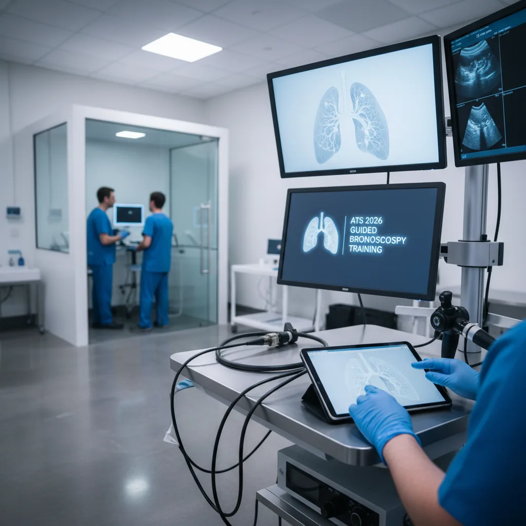
The ATS 2026 session, titled 'A Comprehensive, Hands-On Guide to Basic Bronchoscopy, EBUS, and Guided Bronchoscopy,' is designed to provide participants with practical experience in these essential procedures. The curriculum will likely cover foundational aspects of flexible bronchoscopy, including airway anatomy, instrument handling, and basic biopsy techniques. For EBUS, the focus will be on probe manipulation, identification of anatomical landmarks, and safe execution of TBNA. Participants will learn to differentiate between vascular structures and lymph nodes, a critical skill for preventing complications.4
The guided bronchoscopy component will introduce participants to the principles and practical application of navigation systems. This includes understanding pre-procedural planning using computed tomography (CT) scans, registering the patient's airway to the navigation system, and steering the bronchoscope or guide sheath to peripheral targets. The integration of radial EBUS with guided bronchoscopy will also be emphasized, as it provides real-time confirmation of lesion localization within the peripheral lung parenchyma, thereby maximizing diagnostic accuracy.5
The format of a hands-on guide suggests a strong emphasis on simulation-based training, which has been shown to improve procedural competency and confidence among trainees. Such training environments allow for repeated practice of complex maneuvers and management of potential complications in a controlled setting, without risk to patients.6 The session aims to equip attendees with the practical skills necessary to perform these advanced bronchoscopic procedures effectively and safely in their clinical practice. The absence of specific trial data within the session description itself indicates a focus on skill acquisition and procedural proficiency rather than the presentation of novel research findings. The value lies in its direct applicability to enhancing clinical practice.7
While simulation offers significant advantages, it also presents certain limitations. Simulated environments, despite their sophistication, may not fully replicate the anatomical variability, tissue characteristics, or unexpected challenges encountered in live patients. For instance, the tactile feedback in simulators, while improving, may not perfectly mimic the resistance felt when advancing a needle through different tissue densities or the subtle movements of a patient during a procedure. Furthermore, managing rare but serious complications, such as significant hemorrhage or pneumothorax, in a high-fidelity simulation may not fully prepare a trainee for the acute stress and decision-making required in a real clinical scenario. Therefore, simulation-based training serves as a crucial preparatory step, ideally followed by supervised clinical experience to consolidate skills and adapt to the complexities of patient care. The session's focus on practical application aims to bridge this gap, providing a robust foundation for subsequent clinical exposure.
The ATS 2026 hands-on session on advanced bronchoscopy techniques underscores a critical shift in the expectations for pulmonologists and thoracic surgeons. Competency in EBUS and guided bronchoscopy is no longer a niche skill but a fundamental requirement for optimal patient care in lung disease. Clinicians who do not integrate these techniques into their practice risk providing suboptimal diagnostic pathways, potentially delaying cancer diagnoses or necessitating more invasive procedures. The onus is now on individual practitioners and institutions to ensure adequate training and access to these technologies.
From an industry perspective, the continued emphasis on advanced bronchoscopy highlights the market for sophisticated imaging and navigation systems. Companies like Olympus, Medtronic, and Intuitive Surgical, which develop EBUS scopes, electromagnetic navigation platforms, and robotic bronchoscopy systems, will see sustained demand. However, the cost of these technologies and the associated training can be substantial, posing a barrier for smaller practices or healthcare systems with limited budgets. This disparity could create a two-tiered system of care, where access to cutting-edge diagnostics is not uniformly available.
For patients, the widespread adoption of EBUS and guided bronchoscopy means more accurate and less invasive diagnoses for lung nodules and mediastinal lymphadenopathy. This translates to earlier intervention, potentially improved prognoses for lung cancer, and reduced morbidity associated with surgical biopsies. However, patients in underserved areas or those treated by clinicians without access to these advanced training opportunities may not benefit equally. The medical community must address these access disparities to ensure that advancements in procedural skills translate into equitable improvements in patient outcomes across all demographics.
- The Pivot Advanced bronchoscopic techniques, particularly EBUS and guided bronchoscopy, are now essential for accurate diagnosis and staging of lung diseases.
- The Data While no specific trial data is presented, competency in EBUS has been shown to improve diagnostic yield for mediastinal lymphadenopathy, with reported sensitivities often exceeding 90%.
- The Action Clinicians involved in pulmonary diagnostics should seek training in EBUS and guided bronchoscopy to meet current standards of care and optimize patient outcomes.
ART-2026-118
06/26
Cite This Article
Team TLSFE. Bronchoscopy training: ats 2026 focuses on ebus and guided techniques. The Life Science Feed. Published May 19, 2026. Updated June 28, 2026. Accessed July 4, 2026. https://thelifesciencefeed.com/pulmonology/copd/practice/bronchoscopy-training-ats-2026-focuses-on-ebus-and-guided-techniques.
Editorial & AI Standards
All content is researched from peer-reviewed, open-access sources — published trial data, clinical guidelines, and regulatory filings. AI tools are used solely to structure and summarise that evidence; no AI-generated conclusions appear without editor verification against the primary source.
Every article is reviewed by a named editor before publication. Source citations are listed in the References section. This content does not represent the views of any pharmaceutical company, medical device manufacturer, or healthcare provider.
Licence & Rights
© 2026 The Life Science Feed. All rights reserved. Unless otherwise indicated, all content is the property of The Life Science Feed and may not be reproduced, distributed, or transmitted in any form or by any means without prior written permission.
Medical Disclaimer
The information provided on The Life Science Feed is for educational and informational purposes only. It is not intended as a substitute for professional medical advice, diagnosis, or treatment. Always seek the advice of your physician or other qualified healthcare provider regarding any medical condition or treatment decision. Never disregard professional medical advice or delay in seeking it because of something you have read on this website.
References
1. Ernst A, Silvestri GA, Johnstone D. Interventional pulmonology. Chest. 2005;127(5):1822-1836.
2. Navani N, Nankivell M, Lawrence DR, et al. Lung cancer diagnosis and staging by endobronchial ultrasound-guided transbronchial needle aspiration. Am J Respir Crit Care Med. 2011;184(11):1282-1288.
3. Folch EE, Pritchett MA, Nead MA, et al. Electromagnetic navigation bronchoscopy for peripheral pulmonary lesions: One-year results of the prospective, multicenter NAVIGATE study. J Thorac Oncol. 2019;14(3):445-458.
4. Yasufuku K, Chiyo M, Sekine Y, et al. Real-time endobronchial ultrasound-guided transbronchial needle aspiration of mediastinal and hilar lymph nodes. Chest. 2004;126(1):122-128.
5. Wang Memon M, Murgu SD. Radial endobronchial ultrasound for peripheral pulmonary lesions. J Thorac Dis. 2017;9(Suppl 12):S1371-S1380.
6. Colt HG, Crawford SW, Galbraith O 4th. Virtual reality bronchoscopy simulation: a revolution in procedural training. Chest. 2001;120(4):1333-1339.
7. Wahidi MM, Ernst A, Silvestri GA, et al. Interventional pulmonology: a new subspecialty. Chest. 2010;137(5):1001-1002.