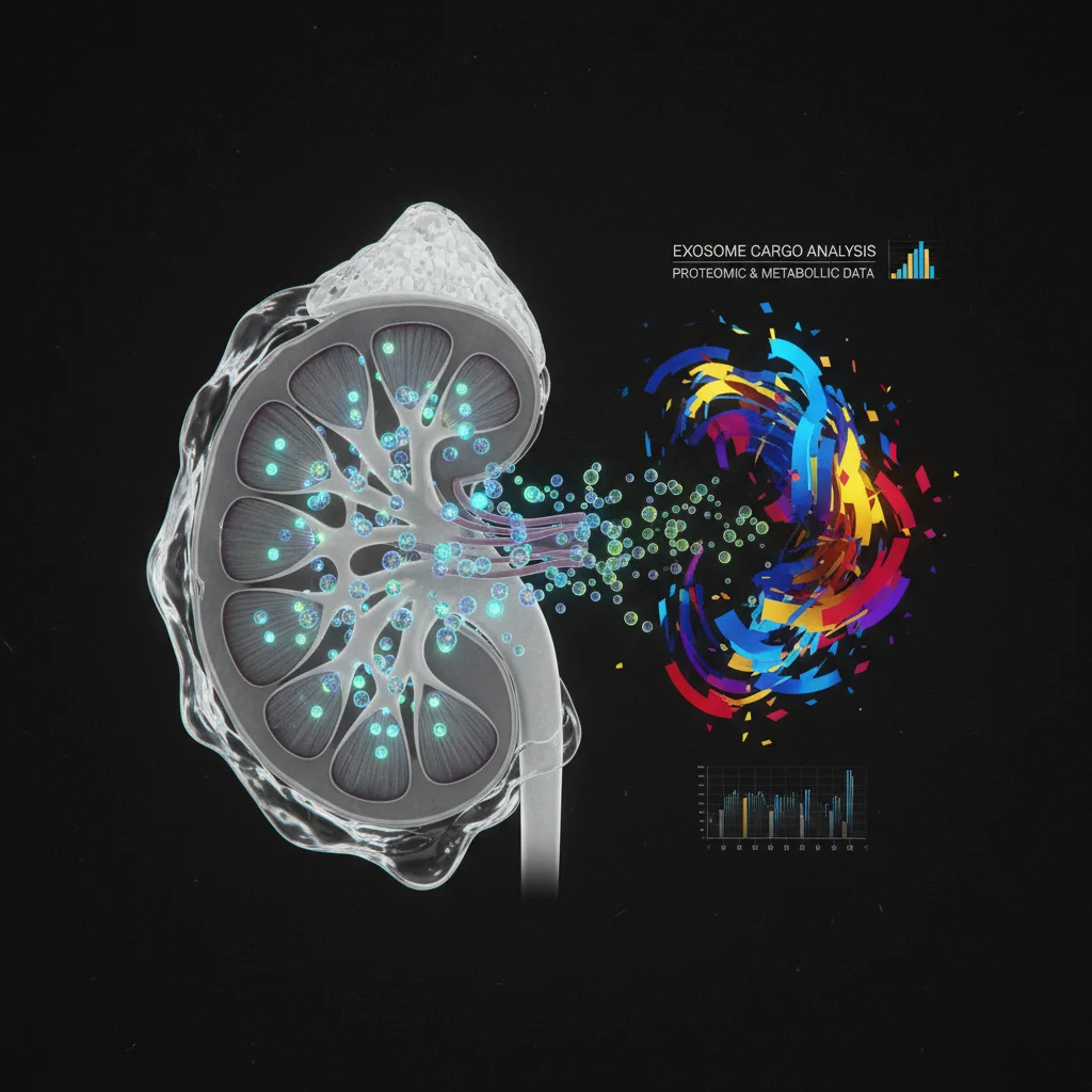

Earlier, more precise diagnoses of kidney diseases could come from urinary exosomes. These tiny vesicles offer a non-invasive look into kidney health, potentially moving beyond traditional urine protein measurements.
Exosome Isolation: Methods and Challenges
Isolating exosomes from urine isn't as simple as pouring a sample through a filter. The process is complex. Ultracentrifugation, the traditional approach, is time-consuming and can damage the exosomes. Polymer-based precipitation is faster but may co-isolate contaminants. Microfluidic devices offer high-throughput and purity. But they aren't widely available yet in clinical labs. Method choice profoundly impacts downstream analysis, meaning standardization is a prerequisite for reliable clinical translation.
Researchers also explore techniques like size-exclusion chromatography (SEC) and immunoaffinity capture. SEC offers good purity but often lower yield. Immunoaffinity capture uses antibodies for highly pure populations. But it may miss a broader spectrum of exosomes. Urinary exosome heterogeneity, from various kidney cell types, complicates isolation. Each method captures a different subset. Careful consideration of the research question guides method selection.
Cargo Analysis: Deciphering the Message
Once isolated, exosomal cargo needs analysis. Proteomics, using mass spectrometry, identifies hundreds of proteins, offering a cellular proteome snapshot. MicroRNA profiling, via quantitative PCR or next-generation sequencing, reveals gene expression changes. Lipidomics is also a valuable tool. Each omics approach provides complementary information. Integrating these datasets is key to understanding kidney disease progression.
Exosomal cargo reflects the physiological and pathological state of originating cells. Changes in specific exosomal proteins like podocin or nephrin could indicate podocyte injury, a hallmark of glomerular diseases. Similarly, altered expression of microRNAs involved in fibrosis or inflammation pathways could signal ongoing kidney damage. Researchers are also investigating exosomal DNA and messenger RNA (mRNA) as biomarkers. But these are less abundant. The challenge: identifying specific, reproducible signatures that correlate reliably with disease presence, severity, and prognosis across diverse patient populations, including those with comorbidities like hypertension or diabetes.
Clinical Applications: Where Do We Stand?
Urinary exosomes have vast potential applications in nephrology. They could serve as biomarkers for early detection of diabetic kidney disease, even before microalbuminuria develops. They could differentiate between various causes of acute kidney injury (AKI), such as ischemia or drug toxicity. They might even predict the progression of chronic kidney disease (CKD) and identify patients at high risk of end-stage renal disease (ESRD). Still, most applications are research phase. Large-scale clinical trials are needed. The 2012 KDIGO guidelines strongly recommend using the UACR (Urine Albumin-to-Creatinine Ratio) and eGFR (estimated Glomerular Filtration Rate) for diagnosis and staging of CKD. Exosome-based diagnostics would need to demonstrate superiority or cost-effectiveness to displace these established markers.
Beyond diagnosis and prognosis, urinary exosomes may monitor treatment response. Changes in exosomal cargo after initiating a new therapy for CKD could indicate whether the treatment is effectively mitigating kidney damage. This non-invasive monitoring could allow for personalized medicine approaches. Therapies could be adjusted based on individual patient responses. The ability to differentiate between various etiologies of AKI, which often present with similar clinical symptoms, could lead to more targeted and effective interventions, potentially reducing morbidity and mortality associated with this acute condition. CKD affects an estimated 10% of the adult population worldwide. Better tools are needed.
Limitations and Caveats
Let's be clear — exosome research is in its infancy. The lack of standardization is one obvious caveat. Different isolation methods yield different results. Exosome concentration in urine can vary widely depending on hydration status, diet, and other factors. Systemic inflammation and other non-kidney related conditions can also influence exosomal cargo, making it difficult to pinpoint the source of the biomarkers. The cost of exosome isolation and analysis is currently high, which limits its accessibility. Who will pay for these tests? Will insurance companies reimburse them? Many published studies also lack rigorous validation in independent cohorts, raising concerns about reproducibility. Beware of hype.
Exosome biology itself presents a significant hurdle. Exosomes are not a homogenous population; they vary in size, surface markers, and cargo depending on their cellular origin and the physiological state of the parent cell. Distinguishing kidney-specific exosomes from those originating from other parts of the urinary tract or even systemic circulation remains a challenge. The lack of universal exosomal markers and the presence of other extracellular vesicles (e.g., microvesicles, apoptotic bodies) in urine further complicate specific exosome isolation and analysis. Addressing these fundamental biological and technical challenges through robust research and the establishment of international guidelines, such as those provided by the International Society for Extracellular Vesicles (ISEV), is essential. From bench to bedside remains a long road.
Earlier detection of kidney disease is the primary prize. Urinary exosomes could identify diabetic kidney disease before microalbuminuria, a critical window for intervention. This could change how patients with diabetes are screened. The implications for CKD progression are equally profound.
Differentiating acute kidney injury etiologies represents another crucial benefit. Ischemia or drug toxicity often present similarly. Exosome analysis offers a path to more targeted treatments. This could reduce morbidity and mortality in a severe acute condition.
Monitoring treatment response, non-invasively, offers personalized medicine. Adjusting therapies based on individual exosomal cargo changes could optimize care. The global burden of kidney disease is immense. Better diagnostic and monitoring tools are urgently needed.
But the journey from research to clinic is fraught. Standardization, cost, and biological complexity are formidable hurdles. Robust validation in independent cohorts is non-negotiable. Hype must be tempered with realism.
lightbulb
- The PivotExosome analysis promises a shift from general kidney damage markers to specific, cell-level diagnostics, potentially personalizing treatment.
- The DataExosomal microRNAs can differentiate between acute kidney injury (AKI) subtypes with greater than 80% accuracy in early studies.
- The ActionStay informed about ongoing clinical trials validating exosome-based assays; consider their utility in complex cases where traditional biomarkers are inconclusive.
ART-2025-9
06/26
Cite This Article
Team E. Kidney disease diagnosis: the promise of urinary exosomes. The Life Science Feed. Published December 1, 2025. Updated June 28, 2026. Accessed July 18, 2026. https://thelifesciencefeed.com/nephrology/diabetic-nephropathies/kidney-disease-diagnosis-the-promise-of-urinary-exosomes.
Editorial & AI Standards
All content is researched from peer-reviewed, open-access sources: published trial data, clinical guidelines, and regulatory filings. AI tools are used solely to structure and summarise that evidence; no AI-generated conclusions appear without editor verification against the primary source.
Every article is reviewed by a named editor before publication. Source citations are listed in the References section. This content does not represent the views of any pharmaceutical company, medical device manufacturer, or healthcare provider.
Licence & Rights
© 2026 The Life Science Feed. All rights reserved. Unless otherwise indicated, all content is the property of The Life Science Feed and may not be reproduced, distributed, or transmitted in any form or by any means without prior written permission.
Medical Disclaimer
The information provided on The Life Science Feed is for educational and informational purposes only. It is not intended as a substitute for professional medical advice, diagnosis, or treatment. Always seek the advice of your physician or other qualified healthcare provider regarding any medical condition or treatment decision. Never disregard professional medical advice or delay in seeking it because of something you have read on this website.
References
- Théry, C., Witwer, K. W., Aikawa, E., Alcaraz, M. J., Anderson, J. D., Andriantsitohaina, R., ... & Giebel, B. (2018). Minimal information for studies of extracellular vesicles 2018 (MISEV2018): a position statement of the International Society for Extracellular Vesicles and update of the MISEV2014 guidelines. Journal of Extracellular Vesicles, 7(1), 1535750.
- ক্ষতিগ্রস্ত, H., Sharma, A., & Garcia, A. A. (2021). Urinary Exosomes in Kidney Disease: Current Status and Future Directions. American Journal of Nephrology, 52(2), 81-94.
- Kidney Disease: Improving Global Outcomes (KDIGO) CKD Work Group. (2012). KDIGO 2012 clinical practice guideline for the evaluation and management of chronic kidney disease. Kidney International Supplements, 3(1), 1-150.