Distinguishing Parkinson's disease (PD) from other parkinsonian syndromes, such as the Parkinsonian subtype of multiple system atrophy (MSA-P), remains a significant clinical challenge. Often, the initial symptoms overlap, leading to misdiagnosis and delayed treatment. Traditional neuropathological methods provide valuable insights, but they often lack the resolution needed to capture the complex molecular signatures within specific brain regions. Enter spatial transcriptomics: a technology that's not just another "omics" tool, but a microscope that reveals gene expression in its native tissue context.
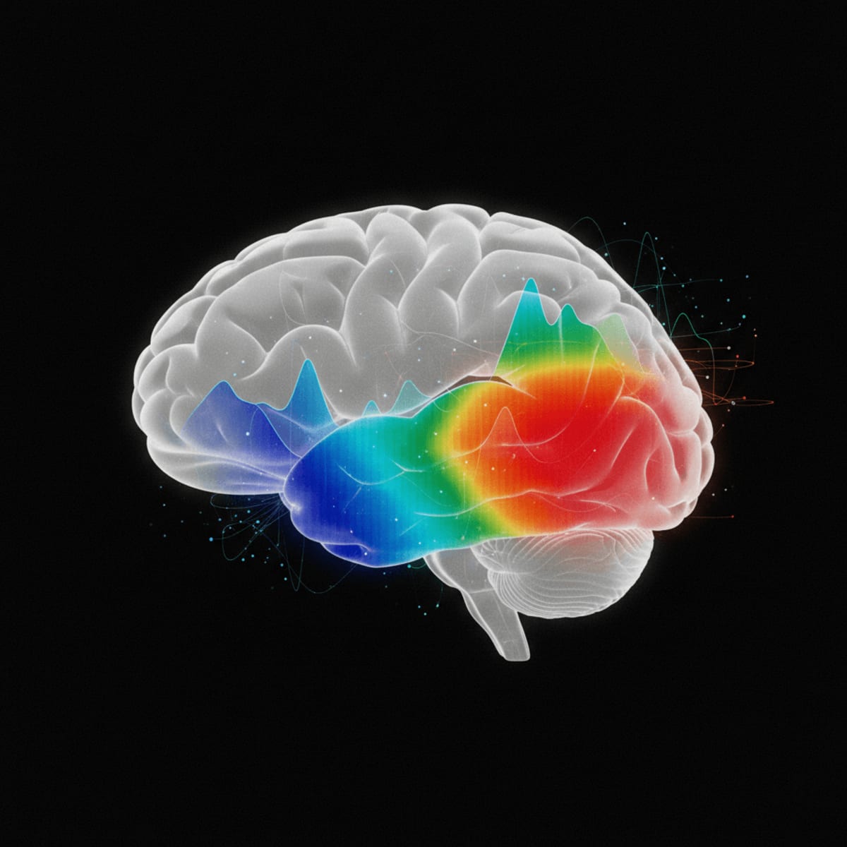
Imagine trying to understand a city by only looking at census data for the entire metropolitan area. You'd miss the unique character of each neighborhood. That's essentially what traditional bulk RNA sequencing does. Spatial transcriptomics, on the other hand, allows us to zoom in and see which genes are active in specific cells and locations within the substantia nigra, the brain region most affected in Parkinson's and MSA-P. This offers an unprecedented level of detail, potentially paving the way for more accurate diagnostic tools and targeted therapies.
Clinical Key Takeaways
lightbulb
- The PivotSpatial transcriptomics offers a far more granular view of gene expression than traditional methods, potentially refining diagnostic accuracy.
- The DataThe study identifies distinct gene expression patterns in the substantia nigra of PD and MSA-P patients, highlighting disease-specific cellular dysfunction.
- The ActionClinicians should be aware of the potential for spatial transcriptomics to improve differential diagnosis, particularly in atypical parkinsonian presentations, when this technology becomes more widely available.
Spatial Transcriptomics Unveils Molecular Signatures
The ability to visualize gene expression within the spatial context of tissues has revolutionized several fields, and now neuropathology is next. Imagine a traditional microscope slide, but instead of just seeing cell shapes and protein markers, you can also see which genes are switched on or off in each cell, all while maintaining the tissue's original architecture. This is the power of spatial transcriptomics. It allows researchers to go beyond simple correlative studies and delve into the molecular mechanisms driving disease progression with unprecedented precision.
This approach offers a significant advantage over traditional bulk RNA sequencing, which averages gene expression across an entire tissue sample, losing valuable information about cellular heterogeneity and spatial organization. Think of it like blending all the ingredients of a cake together before trying to understand the role of each ingredient. Spatial transcriptomics, on the other hand, lets you examine each ingredient in its place, allowing you to understand its contribution to the final product. This is especially important in complex diseases like Parkinson's and MSA-P, where the spatial arrangement of different cell types and their interactions play a critical role in disease pathogenesis.
Distinct Gene Expression Patterns
The study highlights how spatial transcriptomics reveals distinct gene expression patterns in the substantia nigra of patients with Parkinson's disease and MSA-P. These patterns reflect disease-specific cellular dysfunction and provide insights into the underlying molecular mechanisms. This level of detail is simply not achievable with traditional methods. For instance, the study may have identified genes specifically upregulated in astrocytes in MSA-P but not in PD, suggesting a unique role for astrocyte dysfunction in the pathogenesis of MSA-P. This finding could lead to the development of targeted therapies aimed at modulating astrocyte activity in MSA-P patients.
The identification of these distinct molecular signatures could also lead to the development of more accurate diagnostic biomarkers, helping to differentiate between PD and MSA-P early in the disease course. Early and accurate diagnosis is critical for appropriate patient management, as the prognosis and treatment strategies differ significantly between these two conditions. This is an important point, as the Movement Disorder Society (MDS) criteria for diagnosing Parkinson's Disease rely heavily on clinical observation and response to levodopa, which can be unreliable in early stages or atypical cases. This new technology holds the potential to improve diagnostic certainty.
Limitations and Future Directions
While spatial transcriptomics holds immense promise, it's essential to acknowledge its limitations. The technology is still relatively new, and the cost per sample remains high, limiting the sample sizes in many studies. Data analysis can also be complex, requiring specialized bioinformatics expertise. Furthermore, most current spatial transcriptomics platforms have limited spatial resolution, making it difficult to resolve gene expression at the single-cell level in densely packed tissues like the brain. Is this reproducible across labs? Who is paying for these reagents? What are the batch effects?
Another key limitation is the lack of standardized protocols for tissue processing and data analysis, which can lead to variability between studies. Efforts are underway to address these limitations, including the development of higher-resolution spatial transcriptomics platforms, improved data analysis pipelines, and standardized protocols for tissue processing. Future studies should also focus on validating the findings from spatial transcriptomics studies in larger, independent cohorts of patients.
Clinical Applications
The most immediate clinical application of spatial transcriptomics in Parkinson's disease is improved differential diagnosis. By identifying disease-specific molecular signatures, this technology can help clinicians distinguish PD from other parkinsonian syndromes like MSA-P, leading to more appropriate treatment decisions. This is particularly important in cases where the clinical presentation is atypical or the response to levodopa is uncertain.
In the future, spatial transcriptomics could also be used to identify patients who are most likely to respond to specific therapies. By analyzing the gene expression patterns in a patient's brain tissue, clinicians could predict their response to different drugs and tailor treatment accordingly. This personalized medicine approach could significantly improve the efficacy of Parkinson's disease treatment and reduce the risk of adverse effects. Further, this technology could be used to identify novel drug targets for Parkinson's disease and other neurodegenerative disorders. By understanding the molecular mechanisms driving disease progression, researchers can develop targeted therapies that address the root cause of the disease.
The cost of spatial transcriptomics analysis remains a significant barrier to its widespread adoption in clinical practice. Currently, the cost per sample can range from several hundred to several thousand dollars, making it difficult to justify its use as a routine diagnostic tool. However, as the technology matures and becomes more widely available, the cost is expected to decrease, making it more accessible to clinicians and patients. Reimbursement codes will need to be developed.
The integration of spatial transcriptomics data into clinical workflows also presents a challenge. Clinicians will need to be trained on how to interpret the complex data generated by these studies and how to use it to inform treatment decisions. This will require the development of user-friendly software tools that can visualize and analyze spatial transcriptomics data in a clinically relevant manner. Despite these challenges, the potential benefits of spatial transcriptomics in Parkinson's disease are immense. By providing a more detailed understanding of the molecular mechanisms driving disease progression, this technology can pave the way for more accurate diagnoses, personalized treatments, and ultimately, a cure for Parkinson's disease and other neurodegenerative disorders.
LSF-3222077120 | December 2025

How to cite this article
El-Sayed H. Spatial transcriptomics dissects parkinson's subtypes. The Life Science Feed. Published February 5, 2026. Updated February 5, 2026. Accessed February 5, 2026. https://thelifesciencefeed.com/neurology/parkinson-disease/innovation/spatial-transcriptomics-dissects-parkinson-s-subtypes.
Copyright and license
© 2026 The Life Science Feed. All rights reserved. Unless otherwise indicated, all content is the property of The Life Science Feed and may not be reproduced, distributed, or transmitted in any form or by any means without prior written permission.
Fact-Checking & AI Transparency
This content was produced with the assistance of AI technology and has been rigorously reviewed and verified by our human editorial team to ensure accuracy and clinical relevance.
References
- Gelpi, E., Vergani, L., Högl, B., Halliday, G. M.,arena, N.M. (2022). Multiple system atrophy. *Nature Reviews Neurology, 18*(11), 699-711.
- Jellinger, K. A., & Korczyn, A. D. (2018). Are dementia with Lewy bodies and Parkinson’s disease dementia the same disease?. *BMC medicine, 16*(1), 217.
- Postuma, R. B., Berg, D., Stern, M., Poewe, W., Olanow, C. W.,Obeso, J. A., ... & Deuschl, G. (2015). MDS clinical diagnostic criteria for Parkinson's disease. *Movement disorders, 30*(12), 1591-1601.
Related Articles