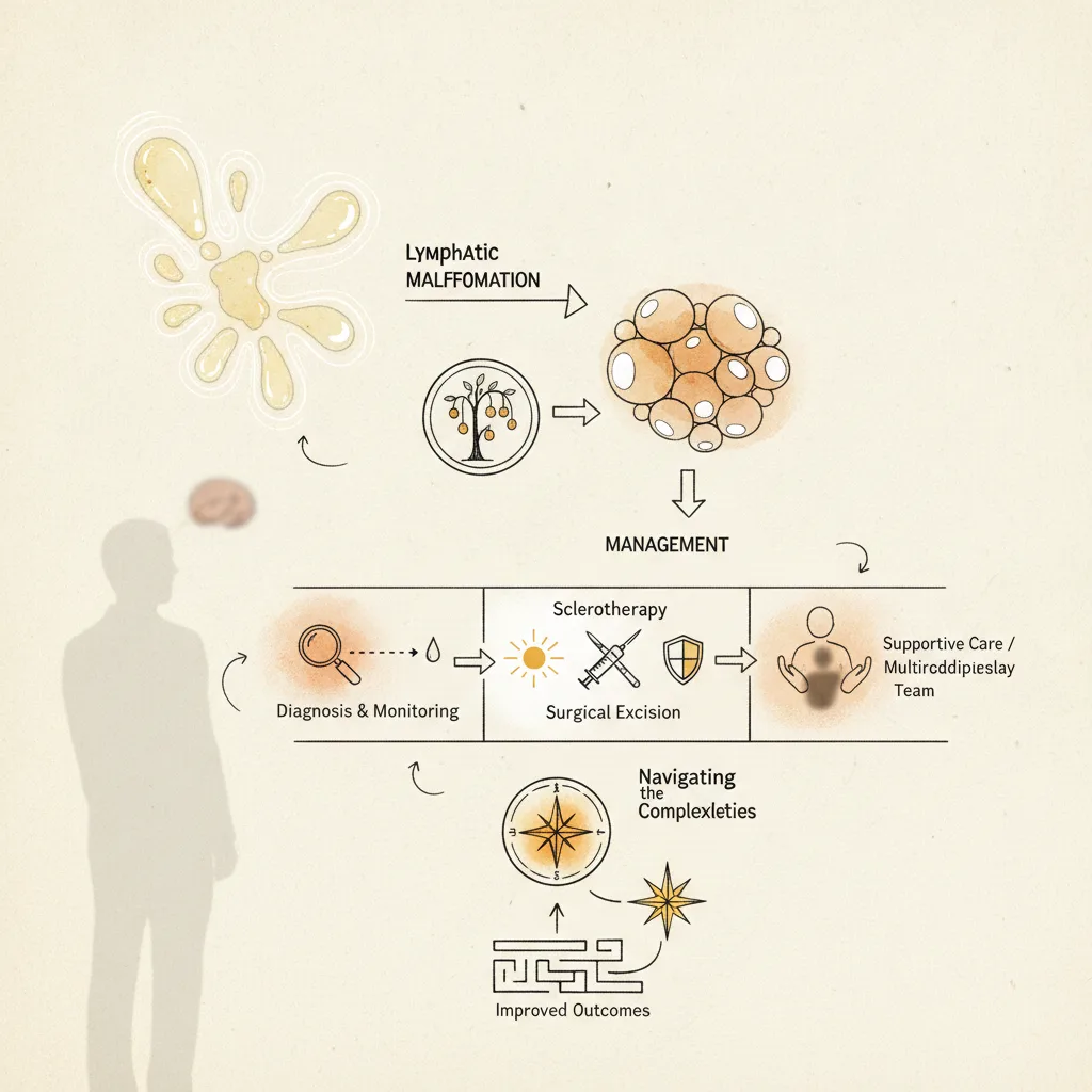

Lymphatic malformations (LMs), often presenting as cystic hygromas, represent a spectrum of congenital anomalies arising from aberrant lymphatic development. These lesions, while benign, can cause significant morbidity due to their location, size, and potential for rapid expansion, posing a persistent challenge for clinicians. Effective management requires a precise understanding of their natural history and the efficacy of available therapeutic strategies, which range from conservative observation to complex surgical and sclerotherapeutic interventions, as detailed in the Medscape premium guide on the topic.
Lymphatic malformations, historically termed cystic hygromas when macrocystic, are congenital anomalies of the lymphatic system. They result from a failure of lymphatic vessels to connect properly with the venous system, leading to fluid accumulation and cyst formation. These lesions are most commonly found in the head and neck region (approximately 75%), but can occur anywhere in the body, including the axilla, mediastinum, and retroperitoneum. Their presentation is highly variable, ranging from small, asymptomatic lesions to large, disfiguring masses that compromise vital structures, necessitating a careful diagnostic and management strategy. The clinical presentation often dictates the urgency and type of intervention required.
The classification of LMs is crucial for guiding treatment. They are broadly categorized into macrocystic (cysts >1 cm), microcystic (cysts <1 cm), or mixed types. Macrocystic lesions are typically amenable to sclerotherapy, while microcystic and mixed lesions often present greater challenges, sometimes requiring surgical excision or alternative therapies. The diagnostic workup typically involves imaging modalities such as ultrasound, MRI, and CT scans to delineate the extent of the lesion, its relationship to adjacent structures, and its internal architecture. MRI is particularly valuable for detailed anatomical mapping and differentiating LMs from other vascular anomalies. The choice of treatment hinges on these imaging characteristics, the lesion's location, and the patient's age and overall health.
The Evolution of Management Strategies
Historically, surgical excision was the primary treatment for lymphatic malformations. The goal was complete removal, but this often proved challenging due to the infiltrative nature of LMs and their close proximity to critical neurovascular structures. Incomplete excision frequently led to recurrence, sometimes with more aggressive growth patterns. Surgical morbidity, including nerve damage, scarring, and lymphatic fistulas, was also a significant concern, particularly for large or deeply invasive lesions. This led to a search for less invasive, yet effective, alternatives that could reduce the burden on patients and improve cosmetic and functional outcomes.
The advent of sclerotherapy marked a significant shift in LM management. This technique involves injecting a sclerosing agent directly into the lymphatic cysts, inducing an inflammatory reaction that leads to fibrosis and collapse of the lymphatic spaces. Various sclerosing agents have been employed, each with its own mechanism of action, efficacy profile, and potential side effects. The choice of agent often depends on the type and location of the LM, as well as the clinician's experience and institutional protocols. Sclerotherapy has largely supplanted surgery as the first-line treatment for many macrocystic LMs, particularly those in anatomically challenging areas.
Sclerotherapy: Agents and Efficacy
Several sclerosing agents have demonstrated efficacy in treating lymphatic malformations. Doxycycline, a tetracycline antibiotic, is widely used due to its availability, relatively low cost, and favorable safety profile. It induces a sterile inflammatory reaction, leading to fibrosis and obliteration of the lymphatic channels. Studies show doxycycline achieves complete or near-complete resolution in approximately 60-70% of macrocystic LMs after multiple injections. The main side effects include localized pain, swelling, and skin discoloration, which are generally transient. For lesions in the head and neck, careful dosing and injection technique are paramount to avoid complications such as nerve irritation or airway compromise.
OK-432 (Picibanil), a lyophilized preparation of a low-virulence strain of Streptococcus pyogenes, is another well-established sclerosing agent, particularly popular in Japan and parts of Europe. It works by stimulating a localized immune response, leading to inflammation and subsequent fibrosis. OK-432 demonstrates high efficacy, with reported complete or near-complete resolution rates of 70-80% for macrocystic LMs, often requiring fewer treatment sessions than doxycycline. Systemic side effects, such as fever, malaise, and leukocytosis, are more common with OK-432 but are typically self-limiting and manageable with antipyretics. Local reactions include swelling and tenderness, which can be significant but usually resolve within a few days. The mechanism of action, involving cytokine release and macrophage activation, makes it particularly effective for lesions with a significant inflammatory component.
Other sclerosing agents include bleomycin, an antineoplastic antibiotic, which has shown efficacy in both macrocystic and microcystic LMs. Bleomycin induces DNA strand breaks, leading to cellular damage and fibrosis. Its use is often reserved for more refractory cases or those with a significant microcystic component where other agents have failed. Efficacy rates for bleomycin range from 50-70%, but concerns about potential systemic toxicity, including pulmonary fibrosis, necessitate careful monitoring, especially with repeated treatments or large doses. The risk of pulmonary toxicity is dose-dependent, making it less suitable for very large or diffuse lesions requiring high cumulative doses.
Ethanol (absolute alcohol) is a potent sclerosing agent that causes protein denaturation and cellular necrosis, leading to rapid fibrosis. It is highly effective, with resolution rates up to 80-90% in selected macrocystic LMs. But its use is limited by significant risks, including severe pain, skin necrosis, nerve damage, and systemic toxicity (e.g., cardiac depression, respiratory arrest) if absorbed systemically. Therefore, absolute ethanol sclerotherapy is typically performed under general anesthesia with strict volume control and careful monitoring, often reserved for lesions that are unresponsive to less toxic agents or those with a high risk of recurrence. Its rapid action requires precise injection and often fluoroscopic guidance to ensure accurate delivery and minimize extravasation.
Sodium tetradecyl sulfate (STS), a detergent sclerosant, is also used, particularly for smaller, more superficial LMs. It acts by damaging endothelial cells, leading to vessel collapse and fibrosis. STS is generally well-tolerated, with localized pain and swelling as the main side effects. Its efficacy is comparable to doxycycline for smaller macrocystic lesions, but it may be less effective for larger or more complex LMs. The concentration of STS used is critical, as higher concentrations increase efficacy but also the risk of local tissue damage.
Surgical Intervention: When and Why
Despite the advancements in sclerotherapy, surgery retains a critical role in the management of lymphatic malformations. It is often indicated for lesions that are refractory to sclerotherapy, those causing acute airway obstruction or other vital organ compression, or those with significant cosmetic disfigurement that cannot be addressed by less invasive means. Complete surgical excision remains the ideal goal, but partial resection or debulking may be necessary to preserve function or minimize morbidity, especially when lesions infiltrate critical structures. The decision to operate is always a balance between the potential benefits of removal and the risks of surgical complications.
For microcystic or mixed LMs, which often have an infiltrative growth pattern, surgery can be particularly challenging. These lesions are less responsive to sclerotherapy due to their small, interconnected lymphatic channels. In such cases, surgical debulking aims to reduce mass effect and improve function, even if complete removal is not feasible. Advances in surgical techniques, including microsurgery and the use of intraoperative imaging, have improved outcomes and reduced complications. But recurrence rates for incompletely excised microcystic LMs remain high, underscoring the need for careful patient selection and realistic expectations.
The timing of surgery is also a consideration. For congenital LMs, early intervention may be necessary if there is airway compromise or feeding difficulties. But for stable lesions, deferring surgery until the child is older may allow for better anatomical definition and reduced surgical risk. Multidisciplinary teams, including pediatric surgeons, interventional radiologists, otolaryngologists, and plastic surgeons, are essential for developing individualized treatment plans that optimize outcomes and minimize long-term sequelae. The Oxford Handbook of Paediatrics offers a concise overview of congenital anomalies and their management in children, providing a useful reference for general practitioners and specialists alike.
Emerging Pharmacotherapies and Targeted Approaches
The understanding of the molecular pathogenesis of lymphatic malformations has opened avenues for targeted pharmacotherapy. Research indicates that many LMs are associated with somatic mutations in genes involved in the PI3K/AKT/mTOR pathway, such as PIK3CA. This pathway plays a crucial role in cell growth, proliferation, and angiogenesis. Inhibitors of the mTOR pathway, such as sirolimus (rapamycin), have shown promise in treating refractory or diffuse LMs, particularly those with identified PIK3CA mutations. Sirolimus is an immunosuppressant that inhibits mTOR, thereby reducing cell proliferation and angiogenesis. It has been used off-label for LMs, with case series and small studies reporting reductions in lesion size, pain, and functional impairment. The drug is administered orally, offering a systemic treatment option for widespread or inaccessible lesions.
But sirolimus therapy is not without its challenges. It requires careful monitoring of drug levels and potential side effects, which include immunosuppression, hyperlipidemia, stomatitis, and proteinuria. The optimal dosing, duration of treatment, and long-term efficacy and safety in LM patients are still under investigation. But for patients with extensive, multifocal, or otherwise untreatable LMs, sirolimus represents a significant therapeutic advance, offering a non-invasive option to manage disease progression and symptoms. Its role is evolving, and it is typically considered after conventional sclerotherapy and surgery have been exhausted or deemed unsuitable.
Other targeted therapies are also under investigation. For example, agents targeting VEGF (vascular endothelial growth factor) pathways, which are involved in lymphangiogenesis, may hold future potential. But these are largely experimental and not yet part of standard clinical practice. The complexity of LM pathophysiology suggests that a personalized medicine approach, guided by genetic profiling of the lesion, may become increasingly important in selecting the most effective therapy.
Complications and Long-Term Management
Lymphatic malformations can lead to a range of complications, both acute and chronic. Acute complications include infection (lymphangitis or cellulitis), hemorrhage into the cysts, and rapid expansion causing airway obstruction or nerve compression. Infections often require antibiotic therapy and sometimes drainage. Hemorrhage can cause sudden swelling and pain, and may necessitate urgent intervention. Chronic complications include cosmetic disfigurement, functional impairment (e.g., dysphagia, speech difficulties, limb swelling), and recurrent infections. The management of these complications is integral to the overall care plan.
Long-term management of LMs often involves a combination of surveillance, supportive care, and repeat interventions as needed. Even after successful treatment, recurrence is possible, particularly for microcystic or mixed lesions. Regular follow-up with imaging is essential to monitor for recurrence or progression. Physical therapy and occupational therapy may be necessary to address functional deficits, especially in cases affecting the limbs or joints. Psychological support is also important for patients and families coping with the chronic nature and potential disfigurement associated with LMs. The goal is not just to eradicate the lesion, but to optimize the patient's quality of life.
The open-label nature of many sclerotherapy studies is an obvious caveat. While efficacy rates are reported, the lack of placebo-controlled trials makes it difficult to definitively quantify the true benefit compared to natural regression or observation alone, particularly for smaller, less symptomatic lesions. But for larger, symptomatic LMs, the clinical improvement observed after sclerotherapy is often dramatic and clearly attributable to the intervention. The heterogeneity of LMs, both in terms of their anatomical location and histological subtype, also complicates direct comparisons between studies and treatment modalities. A lesion in the neck behaves differently from one in the axilla, and a macrocystic lesion responds differently from a microcystic one. These intrinsic differences make it challenging to establish universal treatment algorithms.
The long-term safety data for some sclerosing agents, particularly in pediatric populations, also requires ongoing scrutiny. While agents like doxycycline and OK-432 have generally favorable safety profiles, the cumulative effects of multiple treatments over many years are not fully understood. The potential for long-term scarring, tissue atrophy, or functional impairment, even with successful resolution, must be considered. For systemic therapies like sirolimus, the long-term impact of chronic immunosuppression and metabolic side effects in children is a significant concern, necessitating careful risk-benefit assessment for each patient. The trial was not powered to detect differences in rare complications, and that gap matters for agents with known systemic toxicities. Lymphatic malformation therapy was tested only in symptomatic patients; whether benefits extend to asymptomatic lesions remains unclear, and observation remains a valid strategy for many stable, non-critical lesions.
The shift from radical surgery to targeted sclerotherapy for lymphatic malformations represents a significant improvement in patient care, particularly for macrocystic lesions. Clinicians should prioritize sclerotherapy with agents like doxycycline or OK-432 as a primary intervention, leveraging their efficacy and generally favorable safety profiles. This approach minimizes surgical morbidity and often achieves excellent cosmetic and functional outcomes, especially in anatomically sensitive areas.
But the persistent challenges with microcystic and mixed lesions, and those refractory to sclerotherapy, underscore the need for a multidisciplinary approach. Integrating interventional radiology, pediatric surgery, and, increasingly, medical oncology for targeted pharmacotherapies like sirolimus, is essential. The complexity of these cases demands collaborative decision-making to tailor treatment to the specific lesion characteristics and patient needs.
The emergence of mTOR inhibitors like sirolimus for LMs with PIK3CA mutations offers a systemic option for widespread or surgically inaccessible disease. This development highlights the growing importance of genetic profiling in guiding therapy, moving towards a more personalized medicine paradigm. But the long-term safety and optimal duration of sirolimus in pediatric populations require further robust investigation before it becomes a routine first-line therapy.
Ultimately, the goal remains to improve patient quality of life while minimizing treatment-related morbidity. For many LMs, conservative observation remains a valid initial strategy, particularly for small, asymptomatic lesions. Intervention should be reserved for those causing symptoms, functional impairment, or significant cosmetic concerns, always balancing the potential benefits against the risks of treatment.
- The Pivot Management strategies for lymphatic malformations have evolved from primarily surgical to a multimodal approach incorporating sclerotherapy and targeted pharmacotherapy.
- The Data Sclerotherapy with agents like doxycycline or OK-432 achieves complete or near-complete resolution in 60-80% of macrocystic LMs, but efficacy varies significantly by lesion type.
- The Action Clinicians should consider sclerotherapy as a first-line intervention for accessible macrocystic LMs, reserving surgery for refractory cases or those with critical anatomical impingement.
ART-2026-994
·07/26
Drafted with AI assistance, reviewed and approved by the editorial team. This publication is intended for healthcare professionals, researchers, and life science industry professionals. Content is provided for informational and educational purposes only and does not constitute medical advice.

I specialise in clinical trial methodology and drug development, from Phase I to post-approval. My reports cover what got studied, what did not, and why. Based in Boston, reporting globally.
Cite This Article
Prescott D, Lopes W. Lymphatic malformations: is your management approach outdated?. The Life Science Feed. Published July 29, 2026. Updated July 29, 2026. Accessed July 29, 2026. https://thelifesciencefeed.com/pediatrics/adhd/practice/lymphatic-malformations-is-your-management-approach-outdated.
Editorial & AI Standards
All content is researched from peer-reviewed, open-access sources: published trial data, clinical guidelines, and regulatory filings. AI tools are used solely to structure and summarise that evidence; no AI-generated conclusions appear without editor verification against the primary source.
Every article is reviewed by a named editor before publication. Source citations are listed in the References section. This content does not represent the views of any pharmaceutical company, medical device manufacturer, or healthcare provider.
Licence & Rights
© 2026 The Life Science Feed. All rights reserved. Unless otherwise indicated, all content is the property of The Life Science Feed and may not be reproduced, distributed, or transmitted in any form or by any means without prior written permission.
Medical Disclaimer
The information provided on The Life Science Feed is for educational and informational purposes only. It is not intended as a substitute for professional medical advice, diagnosis, or treatment. Always seek the advice of your physician or other qualified healthcare provider regarding any medical condition or treatment decision. Never disregard professional medical advice or delay in seeking it because of something you have read on this website.
References
1. Mäkinen T, Boon LM, Vikkula M, Alitalo K. Lymphatic Malformations: Genetics, Mechanisms and Therapeutic Strategies. Circ Res. 2021;129(1):136-154. doi:10.1161/CIRCRESAHA.121.318142
2. Pessanha I, Bravo M, Piedade C, Lopes MF. Complex lymphatic malformations in pediatrics: a review of treatment options. Minerva Pediatr (Torino). 2022;74(1):70-80. doi:10.23736/S2724-5276.20.06037-5
3. Kulungowski AM, Patel M. Lymphatic malformations. Semin Pediatr Surg. 2020;29(5):150971. doi:10.1016/j.sempedsurg.2020.150971
4. Kalwani NM, Rockson SG. Management of lymphatic vascular malformations: A systematic review of the literature. J Vasc Surg Venous Lymphat Disord. 2021;9(4):1077-1082. doi:10.1016/j.jvsv.2021.01.013
5. Wiegand S, Wichmann G, Dietz A. Treatment of Lymphatic Malformations with the mTOR Inhibitor Sirolimus: A Systematic Review. Lymphat Res Biol. 2018;16(4):330-339. doi:10.1089/lrb.2017.0062
6. De Maria L, De Sanctis P, Balakrishnan K, Tollefson M, Brinjikji W. Sclerotherapy for lymphatic malformations of head and neck: Systematic review and meta-analysis. J Vasc Surg Venous Lymphat Disord. 2020;8(1):154-164. doi:10.1016/j.jvsv.2019.09.007