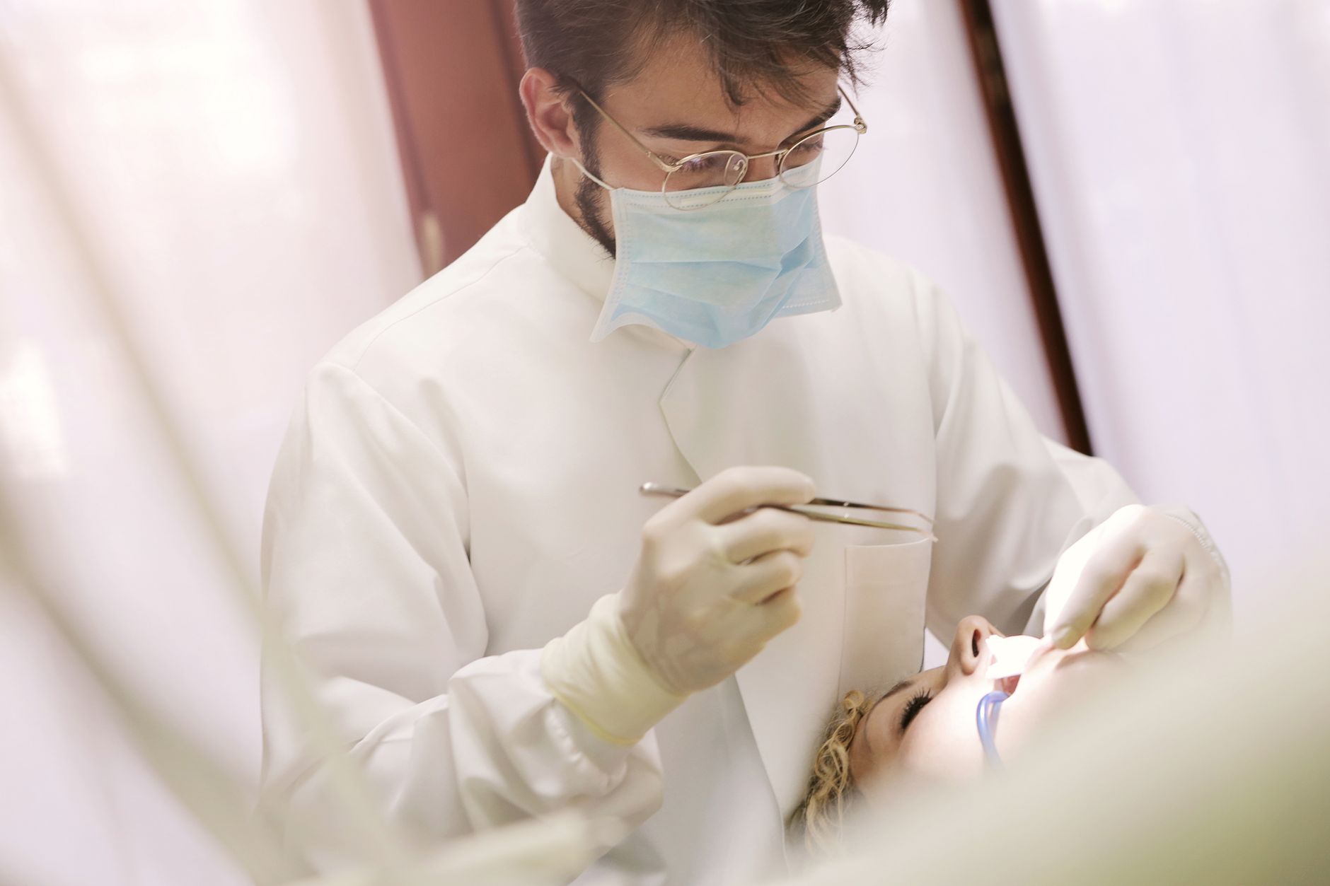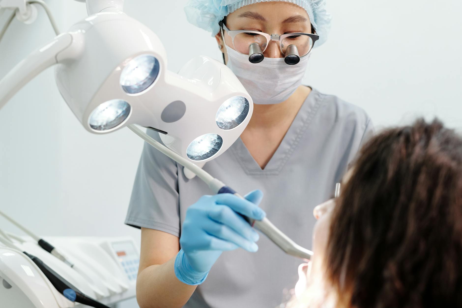

Dental trauma in children, particularly injuries affecting permanent teeth with immature apices, demands a careful approach to treatment. The primary goal extends beyond merely preserving the tooth; it involves fostering continued root development and maintaining pulp vitality, which is important for long-term prognosis. Current guidance attempts to balance these complex objectives, but the practical application often requires significant clinical judgment.
Traumatic dental injuries in the developing dentition are a common occurrence, often leading to significant distress for both children and their parents. The immature apex, characterized by an open foramen and thin dentinal walls, presents a biological opportunity for continued root maturation, but also a vulnerability. Preserving the vitality of the dental pulp is paramount, as it dictates the potential for apexogenesis, the physiological process of root development and apical closure. When pulp vitality is compromised, the challenge shifts to inducing apexification, the formation of a calcified barrier at the root end, or regenerating the pulp-dentin complex.
The management of these injuries is highly dependent on the type of trauma, the extent of pulp involvement, and the stage of root development. Clinicians must consider everything from simple enamel fractures to complex avulsions. The Oxford Handbook of Paediatrics offers a concise overview of the initial assessment, but detailed dental trauma guidance provides the specific protocols. A thorough clinical and radiographic examination is always the starting point, establishing a baseline for pulp sensibility testing and assessing for displacement or fracture.
Pulp Exposure and Vital Pulp Therapy
For injuries involving pulp exposure, such as complicated crown fractures, the immediate concern is preventing bacterial contamination and preserving the remaining vital pulp tissue. Direct pulp capping or partial pulpotomy are the preferred vital pulp therapies for immature permanent teeth. These procedures aim to maintain the vitality of the radicular pulp, allowing for continued root development. The success of these interventions is highly dependent on the size of the exposure, the time elapsed since the injury, and the absence of irreversible pulpitis.
A partial pulpotomy, where only a small portion of the coronal pulp is removed, is often favored over direct pulp capping for larger exposures or when there is a delay in treatment. This approach removes the inflamed or contaminated superficial pulp tissue while preserving the deeper, healthier radicular pulp. The cavity is then sealed with a biocompatible material, typically calcium hydroxide or mineral trioxide aggregate (MTA), followed by a restorative material. The goal is to create an environment conducive to dentin bridge formation and continued root development.
Revascularization and Regenerative Endodontic Procedures
When the pulp becomes necrotic in an immature permanent tooth, the traditional approach has been apexification, often involving multiple appointments and the placement of calcium hydroxide over several months to induce an apical barrier. But, this method can lead to weakened root structures due to continued thin dentinal walls, making the tooth susceptible to fracture. The field has moved towards regenerative endodontic procedures (REPs), or revascularization, as a more biologically driven alternative.
REPs aim to regenerate a functional pulp-dentin complex within the root canal space. This involves disinfecting the canal system, inducing bleeding into the canal to form a blood clot, and then sealing the tooth with a restorative material. The induced blood clot acts as a scaffold for the ingrowth of new tissue, which can include pulp-like tissue, cementum, and bone. The success of revascularization is measured by increased root length and wall thickness, as well as apical closure, indicating continued development of the root. This approach represents a significant shift from merely closing the apex to actively promoting the maturation of the root structure.
Displacement Injuries and Their Management
Displacement injuries, such as luxations and avulsions, present their own set of challenges for immature teeth. Lateral luxation, extrusive luxation, and intrusive luxation all require careful repositioning and stabilization. The prognosis for pulp survival in these cases is inversely related to the severity of the displacement. For instance, intrusive luxation, where the tooth is driven into the alveolar bone, carries a high risk of pulp necrosis and root resorption, particularly in mature teeth. But, in immature teeth, the wider apical foramen may allow for better revascularization, improving the prognosis for pulp vitality.
Avulsion, the complete displacement of a tooth from its socket, is perhaps the most severe form of dental trauma. The immediate management of an avulsed immature permanent tooth is important for success. Reimplantation as quickly as possible, ideally within 60 minutes, is the single most important factor for success. The storage medium for the tooth prior to reimplantation also plays a major role in the tooth's survival; milk, saline, or saliva are preferred over dry storage. After reimplantation, the tooth is splinted, and systemic antibiotics may be prescribed. The long-term prognosis is often complicated by external root resorption, but revascularization attempts are still a viable option for immature teeth.
Fractures and Their Impact on Development
Root fractures in immature teeth are relatively rare but pose a significant challenge. The location of the fracture, whether in the cervical, middle, or apical third, dictates the treatment approach and prognosis. Horizontal root fractures, if located in the apical or middle third, may heal with calcific callus formation, especially if the coronal fragment is stable and the pulp remains vital. However, if the fracture involves the cervical third, the prognosis is often poorer due to increased mobility and difficulty in achieving stable healing.
Crown-root fractures, which extend below the gingival margin, often require complex restorative solutions or even surgical intervention to expose the fracture line for adequate restoration. The goal is always to preserve as much tooth structure as possible and to maintain pulp vitality, particularly in immature teeth where continued root development is desired. The decision to extract or attempt preservation is a critical one, weighing the long-term prognosis against the complexity and invasiveness of the treatment. For a broader understanding of complex surgical considerations, our coverage on IVUS guidance in PCI outcomes highlights how imaging can inform intricate procedures, a principle that, while distinct, echoes the need for precise diagnostics in dental trauma.
The Role of Follow-Up and Monitoring
Regardless of the initial treatment, meticulous follow-up and monitoring are essential for immature permanent teeth that have experienced trauma. Regular clinical and radiographic examinations are necessary to assess pulp vitality, monitor for signs of root resorption, and evaluate continued root development. Pulp sensibility tests, such as electric pulp testing or cold testing, are used to assess the vitality of the pulp, although their reliability can be variable in recently traumatized teeth. Radiographic evaluation helps to detect apical closure, increased root length and wall thickness, and any pathological changes such as inflammatory or replacement resorption.
The frequency and duration of follow-up appointments vary depending on the type and severity of the injury, but typically extend for several years, until root development is complete or a definitive outcome is established. Early detection of complications, such as pulp necrosis or root resorption, allows for timely intervention and can significantly improve the long-term prognosis of the traumatized tooth. The open-label nature of most dental trauma management, where blinding is impossible, is an obvious caveat in assessing outcomes, but the clinical imperative remains clear.
The emphasis on vital pulp therapy and regenerative endodontic procedures for immature teeth after trauma is a welcome evolution. Moving beyond mere apical closure to actively promoting root development offers a better long-term prognosis, reducing the likelihood of future fractures and improving the overall structural integrity of the tooth. This shift demands that general practitioners and pediatric dentists stay current with techniques that were once considered experimental but are now becoming standard.
Clinicians must recognize the critical window for intervention, particularly in avulsion injuries. The difference between immediate reimplantation and even a short delay can dramatically alter the outcome, often dictating whether a tooth survives or succumbs to resorption. This highlights the need for clear, accessible patient education for parents and caregivers on initial first aid for dental trauma.
The ongoing challenge lies in the variability of outcomes and the need for prolonged follow-up. While regenerative procedures hold promise, they are not universally successful, and the clinician must be prepared to manage complications. The commitment to long-term monitoring is non-negotiable, ensuring that any signs of failure or ongoing pathology are addressed promptly to salvage the tooth or plan for appropriate alternatives.
- The Pivot Contemporary guidance prioritizes pulp preservation and continued root development in immature permanent teeth after trauma.
- The Data Treatment decisions hinge on factors like pulp exposure, stage of root development, and time since injury.
- The Action Clinicians should meticulously assess pulp status and choose interventions that support apexogenesis or apexification, adapting to the specific injury type.
ART-2026-1395
·10/26
Drafted with AI assistance, reviewed and approved by the editorial team. This publication is intended for healthcare professionals, researchers, and life science industry professionals. Content is provided for informational and educational purposes only and does not constitute medical advice.

Thirty years in health journalism, the last fifteen in life sciences. I have reported from every major medical congress and watched blockbuster drugs get revised after approval. I cover what the data says.
Cite This Article
Carter J, Voss M. Navigating dental trauma in children: how guidelines handle the immature apex. The Life Science Feed. Published August 27, 2026. Updated October 4, 2026. Accessed October 11, 2026. https://thelifesciencefeed.com/dentistry/dental-trauma/guidelines/dental-trauma-immature-apex-guidance.
Editorial & AI Standards
All content is researched from peer-reviewed, open-access sources: published trial data, clinical guidelines, and regulatory filings. AI tools are used solely to structure and summarise that evidence; no AI-generated conclusions appear without editor verification against the primary source.
Every article is reviewed by a named editor before publication. Source citations are listed in the References section. This content does not represent the views of any pharmaceutical company, medical device manufacturer, or healthcare provider.
Licence & Rights
© 2026 The Life Science Feed. All rights reserved. Unless otherwise indicated, all content is the property of The Life Science Feed and may not be reproduced, distributed, or transmitted in any form or by any means without prior written permission.
Medical Disclaimer
The information provided on The Life Science Feed is for educational and informational purposes only. It is not intended as a substitute for professional medical advice, diagnosis, or treatment. Always seek the advice of your physician or other qualified healthcare provider regarding any medical condition or treatment decision. Never disregard professional medical advice or delay in seeking it because of something you have read on this website.
References
1. Adserias-Garriga J. A review of forensic analysis of dental and maxillofacial skeletal trauma. Forensic Sci Int. 2019;299:80-88. doi:10.1016/j.forsciint.2019.03.027
2. Lam R. Epidemiology and outcomes of traumatic dental injuries: a review of the literature. Aust Dent J. 2016;61 Suppl 1:4-20. doi:10.1111/adj.12395
3. Laforgia A, Inchingolo AM, Inchingolo F, et al. Paediatric dental trauma: insights from epidemiological studies and management recommendations. BMC Oral Health. 2025;25(1):6. doi:10.1186/s12903-024-05222-5
4. de Lima LGH, Dos Santos CS, Rocha JS, Tanaka O, Rosa EAR, Gasparello GG. Comparative analysis of dental trauma in contact and non-contact sports: A systematic review. Dent Traumatol. 2024;40(5):499-510. doi:10.1111/edt.12959
5. Buser D, Urban I, Monje A, Kunrath MF, Dahlin C. Guided bone regeneration in implant dentistry: Basic principle, progress over 35 years, and recent research activities. Periodontol 2000. 2023;93(1):9-25. doi:10.1111/prd.12539
6. Bastone EB, Freer TJ, McNamara JR. Epidemiology of dental trauma: a review of the literature. Aust Dent J. 2000;45(1):2-9. doi:10.1111/j.1834-7819.2000.tb00234.x