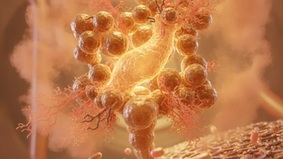
Caveolae, those small, flask-shaped invaginations of the plasma membrane, have long fascinated cell biologists and clinicians alike. Their roles in endocytosis, signal transduction, and lipid homeostasis are well-established, but the precise biophysical mechanisms governing their stability and localisation have remained somewhat elusive. A key insight into this fundamental cellular architecture comes from research published in eLife, which meticulously details how lipid packing contributes to the confinement of caveolae to the plasma membrane.1
Understanding these mechanisms is not merely an academic exercise; caveolae dysfunction is implicated in a range of pathologies, from muscular dystrophies to metabolic disorders and cardiovascular disease. The stability of these structures, therefore, directly impacts cellular health and systemic physiology. This work clarifies a critical biophysical determinant of caveolae integrity.
Caveolae are ubiquitous features of the plasma membrane in many cell types, particularly adipocytes, endothelial cells, and muscle cells. These structures are defined by the presence of caveolin proteins, primarily caveolin-1 and caveolin-2, which polymerise to form the caveolar coat. For years, the prevailing view emphasised the role of these proteins in shaping and stabilising the caveolae. However, the membrane itself, specifically its lipid composition, is increasingly recognised as an active participant in this process. The plasma membrane is not a uniform fluid bilayer; it is a mosaic of distinct lipid domains, often referred to as lipid rafts, which are rich in cholesterol and sphingolipids. These domains are known to be stiffer and more ordered, or 'tightly packed,' than the surrounding membrane.1
Investigators sought to dissect the interplay between caveolin proteins and the surrounding lipid environment, specifically focusing on how lipid packing influences the confinement of caveolae to the plasma membrane. They employed a multi-pronged approach, utilising advanced imaging techniques, biochemical assays, and genetic manipulations in various cell lines, including mouse embryonic fibroblasts (MEFs) and human osteosarcoma (U2OS) cells. The research team systematically altered lipid composition and caveolin expression to observe the resulting effects on caveolae morphology and dynamics.1
Dissecting the membrane's role in caveolae stability
The first step involved establishing a baseline understanding of caveolae dynamics. Researchers observed that caveolae are not static structures; they undergo cycles of budding and retrieval from the plasma membrane. This dynamic behaviour is tightly regulated, and dysregulation can lead to cellular dysfunction. The team hypothesised that increased lipid packing, characteristic of lipid rafts, would stabilise caveolae at the plasma membrane, preventing their internalisation or dissociation.1
To test this, they manipulated the cellular cholesterol content. Cholesterol is a key component of lipid rafts and directly influences membrane fluidity and packing. Depleting cholesterol using methyl-beta-cyclodextrin (MβCD) led to a significant reduction in plasma membrane-localised caveolae. Conversely, enriching cholesterol increased the number of stable caveolae at the cell surface. This indicated a direct correlation between membrane cholesterol levels and caveolae confinement. The effect was dose-dependent, with higher cholesterol concentrations leading to greater stability.1
But cholesterol is not the only player. Sphingolipids, particularly sphingomyelin, also contribute to lipid raft formation and membrane rigidity. The investigators used inhibitors of sphingolipid synthesis, such as myriocin, to reduce cellular sphingomyelin levels. This intervention mirrored the effects of cholesterol depletion, resulting in fewer caveolae confined to the plasma membrane. These findings collectively pointed towards a critical role for tightly packed lipid domains, rich in both cholesterol and sphingolipids, in maintaining caveolae integrity at the cell surface.1
The mechanics of lipid packing and protein interaction
The team then delved into the mechanism by which lipid packing exerts its influence. Caveolin proteins possess a unique hairpin-like structure that inserts into the inner leaflet of the plasma membrane. This insertion is thought to induce membrane curvature, a prerequisite for caveolae formation. The study proposed that the tightly packed lipid environment provides a stable scaffold for these caveolin proteins to assemble and maintain the characteristic flask shape. Without this optimal lipid packing, the membrane might be too fluid or too disordered to support the stable invagination.1
They performed experiments using fluorescent probes that report on membrane order and packing density. These probes consistently showed that caveolae-rich regions of the plasma membrane exhibited higher lipid packing compared to surrounding areas. This was true even in cells where caveolin expression was reduced, suggesting that while caveolins are essential for forming the structure, the lipid environment is crucial for its stability. The researchers also used atomic force microscopy to directly measure membrane stiffness, confirming that caveolae regions were indeed mechanically stiffer.1
Further experiments involved reconstituting caveolin proteins in artificial lipid bilayers with varying lipid compositions. When caveolins were introduced into bilayers mimicking the tightly packed environment of lipid rafts, they readily formed stable, caveolae-like structures. However, in more fluid, less packed lipid environments, caveolin assembly was less efficient, and the resulting structures were less stable and more prone to dissociation. This provided strong evidence that lipid packing is not merely permissive but actively contributes to the structural integrity of caveolae.1
Caveolin-lipid synergy and functional implications
The study also addressed the interplay between caveolin proteins and the lipid environment. While lipid packing is critical, caveolin proteins are indispensable for caveolae formation. Cells lacking caveolin-1, for instance, do not form caveolae, regardless of their lipid composition. But in cells expressing caveolin-1, the lipid environment dictates the efficiency and stability of caveolae formation. This suggests a synergistic relationship: caveolins initiate the curvature, and the tightly packed lipids stabilise it.1
The functional implications of this finding are substantial. Caveolae are known to be involved in mechanosensing, responding to physical forces exerted on the cell membrane. A stiffer, more tightly packed membrane, as suggested by this research, would naturally alter how cells perceive and respond to mechanical stimuli. This could have profound effects on processes like cell migration, tissue development, and even disease progression in conditions such as atherosclerosis, where endothelial cells are constantly subjected to shear stress. The Oxford Handbook of Cardiology details many such conditions where cellular mechanics play a role.
The researchers also explored the impact of lipid packing on caveolae-mediated endocytosis. Caveolae are a major pathway for internalising various molecules, including growth factors, toxins, and even some viruses. When lipid packing was reduced, and caveolae stability compromised, the efficiency of caveolae-mediated endocytosis decreased. This indicates that the structural integrity conferred by lipid packing is directly linked to the functional capacity of these membrane invaginations. This has implications for drug delivery strategies, where understanding the internalisation pathways is paramount.1
Still, the study was primarily conducted in immortalised cell lines, which may not fully recapitulate the complex lipid environment and regulatory mechanisms present in primary tissues in vivo. The precise lipid composition of caveolae can vary significantly between different cell types and physiological states. This variability could introduce nuances not fully captured by the current experimental models. Further research in more physiologically relevant systems, such as primary cell cultures or animal models, would be necessary to fully validate these findings and explore their broader biological context.1
Another consideration is the dynamic nature of lipid packing itself. Membrane lipid composition is not static; it can change rapidly in response to various stimuli, including nutrient availability, stress, and signalling events. The study provides a snapshot of the role of lipid packing, but the temporal regulation of this packing and its impact on caveolae dynamics over time remain areas for further investigation. Understanding how cells actively modulate their membrane lipid packing to control caveolae function could open new avenues for therapeutic intervention.1
The open-label design of some biochemical assays is an obvious caveat, though the biophysical nature of the measurements mitigates some of the typical concerns. The reliance on chemical agents to manipulate lipid composition, while standard, can also introduce off-target effects that might indirectly influence caveolae behaviour. Future studies employing more targeted genetic approaches to alter specific lipid synthesis pathways could provide even cleaner insights into these intricate relationships.1
The study did not explicitly quantify the precise number of caveolae that were internalised versus those that simply disassembled at the plasma membrane. Distinguishing between these two fates is important for fully understanding the dynamic lifecycle of caveolae and how lipid packing influences each step. Advanced live-cell imaging techniques with higher spatial and temporal resolution could help to resolve these fine distinctions.1
This work provides a mechanistic underpinning for how caveolae maintain their structural integrity, moving beyond a purely protein-centric view. Clinicians dealing with conditions like muscular dystrophy, where caveolin mutations lead to unstable caveolae, might consider that membrane lipid composition could be a modifiable factor. Current therapeutic approaches largely focus on gene therapy or protein replacement, but altering the lipid environment could offer a complementary strategy.
The implications extend to metabolic disorders. Caveolae are critical for insulin signalling and lipid uptake in adipocytes. If lipid packing is suboptimal, these processes could be impaired, contributing to insulin resistance or dyslipidaemia. Nutritional interventions or pharmacological agents that subtly modulate membrane lipid composition could, in theory, improve caveolae function and metabolic health.
For cardiovascular specialists, understanding caveolae stability in endothelial cells is paramount. Endothelial caveolae regulate nitric oxide production and mechanotransduction, processes vital for vascular health. Dysfunctional caveolae contribute to atherosclerosis and hypertension. Targeting lipid packing in the endothelium might offer novel avenues for preventing or treating these widespread conditions, moving beyond traditional risk factor management.
The pharmaceutical industry, often focused on receptor-ligand interactions, should take note. Drug delivery strategies that rely on caveolae-mediated endocytosis could be significantly enhanced by optimising the lipid environment of target cells. Designing drugs that subtly alter membrane packing, or co-administering lipid-modulating agents, might improve therapeutic efficacy and reduce off-target effects.
- The Pivot Caveolae stability at the plasma membrane is not solely protein-driven; lipid packing density is a primary determinant.
- The Data Increased lipid packing, specifically through cholesterol and sphingolipid enrichment, directly enhances caveolae confinement.
- The Action Future therapeutic strategies targeting caveolae-related pathologies may need to consider membrane lipid composition alongside protein interactions.
ART-2026-993
·09/26
Drafted with AI assistance, reviewed and approved by the editorial team. This publication is intended for healthcare professionals, researchers, and life science industry professionals. Content is provided for informational and educational purposes only and does not constitute medical advice.

Infectious disease, epidemiology, and global health equity. I have covered outbreaks from Ebola to COVID-19 to mpox. The stories I am most drawn to are where the science is clear and the response is slow.
Cite This Article
Reeves T, Voss M. How lipid packing dictates caveolae stability at the plasma membrane. The Life Science Feed. Published July 27, 2026. Updated September 15, 2026. Accessed September 24, 2026. https://thelifesciencefeed.com/cardiology/hyperlipidemias/research/how-lipid-packing-dictates-caveolae-stability-at-the-plasma-membrane.
Editorial & AI Standards
All content is researched from peer-reviewed, open-access sources: published trial data, clinical guidelines, and regulatory filings. AI tools are used solely to structure and summarise that evidence; no AI-generated conclusions appear without editor verification against the primary source.
Every article is reviewed by a named editor before publication. Source citations are listed in the References section. This content does not represent the views of any pharmaceutical company, medical device manufacturer, or healthcare provider.
Licence & Rights
© 2026 The Life Science Feed. All rights reserved. Unless otherwise indicated, all content is the property of The Life Science Feed and may not be reproduced, distributed, or transmitted in any form or by any means without prior written permission.
Medical Disclaimer
The information provided on The Life Science Feed is for educational and informational purposes only. It is not intended as a substitute for professional medical advice, diagnosis, or treatment. Always seek the advice of your physician or other qualified healthcare provider regarding any medical condition or treatment decision. Never disregard professional medical advice or delay in seeking it because of something you have read on this website.
References
1. eLife. Lipid packing contributes to the confinement of caveolae to the plasma membrane. Accessed May
2024. https://elifesciences.org/articles/108369