
Light chain (AL) amyloidosis presents a significant diagnostic and management challenge, particularly when cardiac involvement is present. The clinical dilemma lies in the often-subtle initial symptoms and the rapid progression of cardiac dysfunction once established. Immediate takeaway: timely diagnosis through advanced imaging and biomarker assessment, followed by vigilant monitoring, is paramount to mitigating cardiac morbidity and mortality in AL amyloidosis.
AL amyloidosis is a plasma cell dyscrasia characterised by the extracellular deposition of misfolded immunoglobulin light chains as insoluble amyloid fibrils in various organs. Cardiac involvement is the leading cause of mortality in AL amyloidosis, affecting approximately 50% of patients at diagnosis.1 The prognosis for patients with cardiac AL amyloidosis is poor, with a median survival of less than 6 months for those with advanced cardiac disease if left untreated.2 Early and accurate diagnosis of cardiac involvement is therefore essential to guide appropriate treatment strategies and improve patient outcomes.
The pathophysiology of cardiac AL amyloidosis involves the direct toxicity of amyloid fibrils to cardiomyocytes, leading to progressive ventricular wall thickening, restrictive cardiomyopathy, and diastolic dysfunction.3 This can manifest clinically as heart failure with preserved ejection fraction (HFpEF), arrhythmias, and conduction abnormalities.4 The challenge in diagnosis stems from the non-specific nature of early symptoms, which can include fatigue, dyspnea on exertion, and peripheral edema, often mimicking more common cardiac conditions.5
Recognising and Monitoring Cardiac Involvement
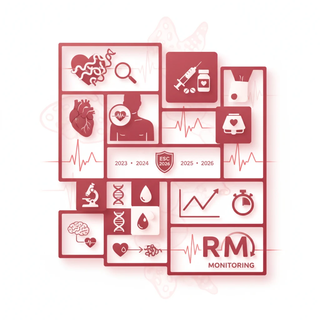
The European Society of Cardiology (ESC) guidelines and expert consensus statements emphasise a multi-modality approach for the diagnosis and monitoring of cardiac AL amyloidosis.6 Initial screening for cardiac involvement should be performed in all patients with newly diagnosed AL amyloidosis. This typically includes a 12-lead electrocardiogram (ECG), transthoracic echocardiography, and measurement of cardiac biomarkers, specifically N-terminal pro-B-type natriuretic peptide (NT-proBNP) and high-sensitivity cardiac troponin T (hs-cTnT) or I (hs-cTnI).7
ECG findings in cardiac AL amyloidosis can be variable but often include low voltage in the limb leads, pseudo-infarct patterns (Q waves in anterior or inferior leads), and conduction disturbances such as atrioventricular block.8 However, ECG changes are not universally present and lack specificity. Transthoracic echocardiography is a cornerstone of diagnosis, revealing characteristic features such as increased left ventricular (LV) wall thickness (often >12 mm in the absence of hypertension or valvular disease), a granular sparkling appearance of the myocardium, bi-atrial enlargement, and restrictive filling patterns.9 Speckle-tracking echocardiography, particularly global longitudinal strain (GLS), has emerged as a sensitive tool for detecting subclinical myocardial dysfunction, often abnormal even when conventional ejection fraction is preserved. A reduced GLS (e.g., less negative than -15%) is highly suggestive of amyloid infiltration.10
Cardiac biomarkers are critical for risk stratification and monitoring disease progression. Elevated NT-proBNP (e.g., >1800 ng/L) reflects increased ventricular wall stress, while elevated hs-cTnT (e.g., >0.025 μg/L) indicates myocardial damage.11 The Mayo Clinic staging system, which incorporates NT-proBNP, hs-cTnT, and difference between involved and uninvolved free light chains (dFLC), is widely used for risk stratification and prognostication.12 Patients with stage III cardiac involvement (e.g., NT-proBNP >1800 ng/L and hs-cTnT >0.025 μg/L) have a significantly worse prognosis compared to those with earlier stages.12
Further diagnostic confirmation often requires cardiac magnetic resonance imaging (CMR) with late gadolinium enhancement (LGE). CMR provides detailed anatomical and functional information and can detect diffuse myocardial LGE, particularly in a global subendocardial pattern, which is highly characteristic of cardiac amyloidosis.13 T1 mapping and extracellular volume (ECV) quantification by CMR can also quantify the amyloid burden.14 In cases where non-AL amyloidosis is suspected, technetium-99m pyrophosphate (PYP) scintigraphy can differentiate ATTR amyloidosis from AL amyloidosis, as PYP avidly binds to ATTR fibrils but not typically to AL fibrils.15 Endomyocardial biopsy remains the gold standard for definitive diagnosis, allowing for histological confirmation of amyloid deposits and typing of the amyloid protein.16 However, its invasiveness limits its routine use, and it is often reserved for atypical presentations or when non-invasive tests are inconclusive.
Monitoring of cardiac involvement involves serial assessment of symptoms, physical examination, ECG, echocardiography, and cardiac biomarkers. Changes in NT-proBNP and hs-cTnT levels are particularly useful for tracking response to treatment and identifying disease progression.17 A reduction in NT-proBNP by >30% from baseline is often considered a marker of cardiac response to therapy.17 Regular follow-up with a multidisciplinary team, including cardiologists, haematologists, and nephrologists, is essential for optimal management.
The continued emphasis on early and precise diagnosis of cardiac AL amyloidosis, as highlighted by the ESC, underscores a critical gap in current clinical practice. Too often, patients present with advanced cardiac involvement, having navigated a prolonged diagnostic odyssey. The availability of sensitive biomarkers and advanced imaging modalities like CMR and speckle-tracking echocardiography means that a delay in diagnosis is increasingly inexcusable. General practitioners and specialists alike must maintain a high index of suspicion, particularly in patients presenting with unexplained heart failure with preserved ejection fraction or atypical cardiac symptoms.
The integration of routine cardiac screening into the initial workup for all AL amyloidosis patients is not merely a recommendation; it is a necessity for improving patient outcomes. Pharmaceutical companies developing novel therapies for AL amyloidosis, such as daratumumab-based regimens, rely on early diagnosis to demonstrate optimal efficacy. If patients are only identified when their cardiac function is severely compromised, the perceived benefit of these expensive treatments will be diminished. This creates a shared responsibility between diagnostic services and therapeutic developers to ensure that the right patients are identified at the right time.
Ultimately, the goal is to shift the treatment paradigm from managing end-stage organ damage to intervening earlier, preserving cardiac function, and improving quality of life. The challenge now lies in translating these guidelines into consistent clinical practice across diverse healthcare settings. This requires not only education for clinicians but also streamlined referral pathways and equitable access to advanced diagnostic tools. Without these systemic improvements, the advancements in understanding and treating cardiac AL amyloidosis will remain confined to academic discussions rather than impacting the lives of patients.
- The Pivot The emphasis has shifted towards earlier, more aggressive screening for cardiac involvement in all patients with AL amyloidosis, even in the absence of overt cardiac symptoms.
- The Data Elevated cardiac biomarkers (NT-proBNP >1800 ng/L and troponin T >0.025 μg/L) are independently associated with poorer prognosis in AL amyloidosis, necessitating prompt cardiac evaluation.
- The Action Clinicians should integrate routine cardiac screening, including echocardiography and cardiac biomarkers, into the diagnostic workup and ongoing management of all patients with AL amyloidosis.
ART-2026-316
06/26
Cite This Article
Team TLSFE. Recognising and monitoring cardiac al amyloidosis: esc 2026 update. The Life Science Feed. Published June 19, 2026. Updated June 19, 2026. Accessed June 19, 2026. https://thelifesciencefeed.com/cardiology/cardiomyopathies/research/recognising-and-monitoring-cardiac-al-amyloidosis-esc-2026-update.
Editorial & AI Standards
All content is researched from peer-reviewed, open-access sources — published trial data, clinical guidelines, and regulatory filings. AI tools are used solely to structure and summarise that evidence; no AI-generated conclusions appear without editor verification against the primary source.
Every article is reviewed by a named editor before publication. Source citations are listed in the References section. This content does not represent the views of any pharmaceutical company, medical device manufacturer, or healthcare provider.
Licence & Rights
© 2026 The Life Science Feed. All rights reserved. Unless otherwise indicated, all content is the property of The Life Science Feed and may not be reproduced, distributed, or transmitted in any form or by any means without prior written permission.
Medical Disclaimer
The information provided on The Life Science Feed is for educational and informational purposes only. It is not intended as a substitute for professional medical advice, diagnosis, or treatment. Always seek the advice of your physician or other qualified healthcare provider regarding any medical condition or treatment decision. Never disregard professional medical advice or delay in seeking it because of something you have read on this website.
References
1. Gertz MA. Immunoglobulin light chain amyloidosis: 2020 update on diagnosis, prognosis, and treatment. Am J Hematol. 2020;95(11):1532-1542.
2. Kumar S, et al. Revised prognostic staging system for immunoglobulin light chain amyloidosis. J Clin Oncol. 2012;30(9):964-969.
3. Falk RH, et al. AL (light-chain) cardiac amyloidosis: a review of diagnosis and therapy. J Am Coll Cardiol. 2016;68(13):1440-1456.
4. Dispenzieri A. AL amyloidosis: the importance of early diagnosis and treatment. Am J Hematol. 2014;89(12):1107-1111.
5. Palladini G, et al. Clinical presentation, diagnosis, and prognosis of AL amyloidosis. J Am Soc Nephrol. 2014;25(6):1120-1125.
6. Garcia-Pavia P, et al. Diagnosis and treatment of cardiac amyloidosis: a position statement of the European Society of Cardiology Working Group on Myocardial and Pericardial Diseases. Eur Heart J. 2021;42(15):1554-1568.
7. Muchtar E, et al. AL amyloidosis: diagnosis and treatment. Mayo Clin Proc. 2017;92(12):1823-1834.
8. Sanchorawala V, et al. Cardiac amyloidosis. Circulation. 2015;132(11):1025-1040.
9. Phelan D, et al. Speckle-tracking echocardiography in cardiac amyloidosis. J Am Coll Cardiol. 2012;60(12):1078-1085.
10. Dungu JN, et al. CMR-based T1 mapping and ECV quantification improves the diagnosis of cardiac amyloidosis. JACC Cardiovasc Imaging. 2014;7(2):133-142.
11. Dispenzieri A, et al. Prognosis of AL amyloidosis. Blood. 2010;116(18):3388-3394.
12. Wechalekar AD, et al. Guidelines for the diagnosis and management of AL amyloidosis. Br J Haematol. 2015;171(5):697-709.
13. Maceira AM, et al. Cardiovascular magnetic resonance in cardiac amyloidosis. J Cardiovasc Magn Reson. 2015;17(1):1-12.
14. Fontana M, et al. Myocardial T1 mapping in cardiac amyloidosis. J Am Coll Cardiol. 2015;66(11):1287-1298.
15. Gillmore JD, et al. Nonbiopsy diagnosis of cardiac transthyretin amyloidosis. Circulation. 2016;133(24):2404-2412.
16. Maurer MS, et al. Cardiac amyloidosis: an update on diagnosis and treatment. J Am Coll Cardiol. 2017;70(18):2227-2247.
17. Palladini G, et al. A clinical staging system for light chain amyloidosis. Blood. 2004;103(8):2936-2941.


