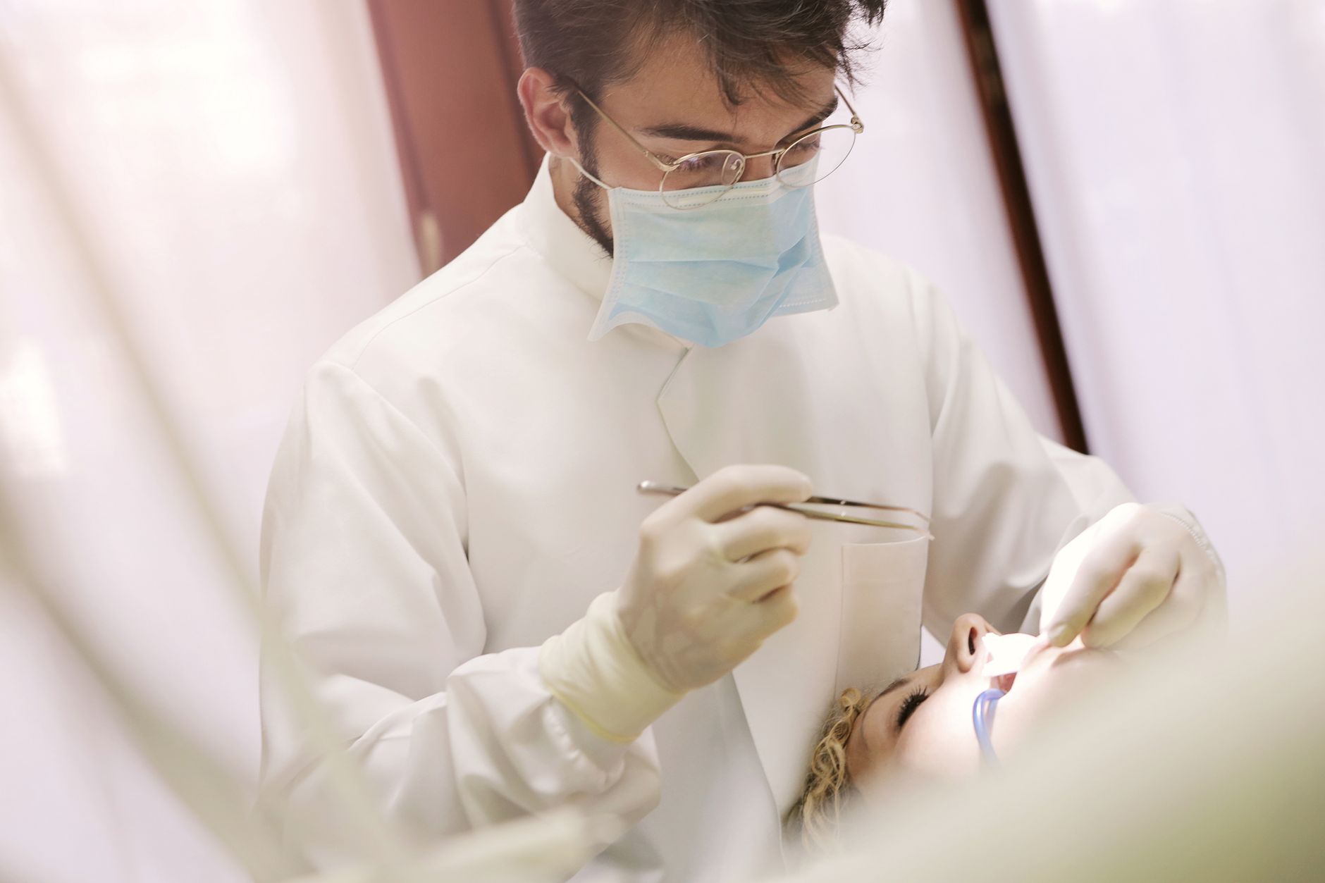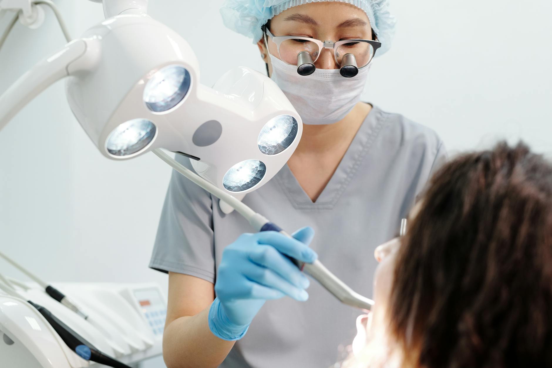

Oral squamous cell carcinoma (OSCC) remains a significant public health burden, with late-stage diagnosis contributing to poor prognoses and high mortality rates. Conventional oral examination (COE) is the standard for detecting suspicious lesions, but its efficacy hinges on clinician experience and the often subtle presentation of early-stage disease. The persistent challenge lies in reliably distinguishing benign lesions from dysplastic or malignant changes at a stage amenable to curative intervention.
Despite the introduction of various adjunctive screening devices, a critical question persists: do these technologies genuinely improve detection rates or diagnostic accuracy beyond what a thorough conventional examination can achieve? A study published in Oral Surgery, Oral Medicine, Oral Pathology and Oral Radiology explored the potential of a novel multimodal optical imaging system to address this gap.1
Oral cancer, particularly OSCC, accounts for a substantial proportion of head and neck cancers, with global incidence rates varying but generally remaining a concern for primary care physicians and specialists alike. Early detection is paramount, as survival rates dramatically improve when the disease is identified at a localised stage. But the visual and tactile nature of conventional oral examination means that even experienced clinicians can miss subtle, early-stage lesions, or conversely, refer benign lesions for unnecessary biopsies. This diagnostic dilemma has driven the development of various adjunctive technologies, from vital tissue staining to autofluorescence imaging, all aiming to enhance the clinician's ability to identify suspicious areas.1
But these adjunctive techniques have consistently struggled to demonstrate a clear, unequivocal advantage over COE in terms of diagnostic accuracy. Many have shown high sensitivity but poor specificity, leading to an increase in false positives and subsequent over-referral for biopsy. Others have failed to achieve sufficient sensitivity to reliably detect all true positives. This lack of superior performance has left clinicians without a definitive tool to supplement their standard examination protocols, underscoring the ongoing need for more effective screening modalities. The primary care assessment of oral ulcers and patches remains a critical skill, often without the benefit of advanced diagnostics.1
The Unmet Need in Oral Cancer Screening
The current market of oral cancer screening relies heavily on the subjective interpretation of visual and tactile cues during a conventional oral examination. This method, while fundamental, is inherently limited by the human eye's ability to discern subtle changes in tissue morphology and the clinician's experience. Early dysplastic lesions or very early carcinomas often present as innocuous-looking white or red patches, or even as seemingly normal mucosa, making definitive identification challenging without invasive biopsy. The goal of any adjunctive device is to provide objective, non-invasive information that can guide the clinician towards the most suspicious areas, thereby improving the yield of biopsies and reducing the number of unnecessary procedures.1
Existing adjunctive screening devices, such as those employing autofluorescence or chemiluminescence, have been marketed with the promise of enhancing lesion visualisation. But a comprehensive review of the literature reveals that these tools have not consistently delivered on this promise in clinical settings. Many studies report high sensitivity, meaning they correctly identify a large proportion of true positives, but their specificity often falls short. This leads to a significant number of false-positive results, where benign lesions are flagged as suspicious, causing patient anxiety and increasing healthcare costs due to unnecessary referrals and biopsies. Conversely, some devices, while specific, lack the sensitivity to reliably detect all precancerous or cancerous lesions, leaving clinicians with a false sense of security.1
A Novel Approach to Optical Imaging
Malik, Jabbour, and Cheng developed a novel multimodal optical imaging system designed to overcome the limitations of previous adjunctive devices.1 Their system integrates two distinct optical imaging techniques: macroscopic biochemical imaging of fluorescence lifetime imaging (FLIm) and subcellular morphologic imaging of reflectance confocal microscopy (RCM). The rationale behind this multimodal approach is to leverage the strengths of each technique, providing a more comprehensive assessment of tissue changes indicative of early oral cancer. FLIm offers insights into the biochemical composition of tissues by measuring the decay rate of endogenous fluorophores, which can change in the presence of dysplasia or malignancy. RCM, on the other hand, provides high-resolution, real-time images of cellular and subcellular structures, allowing for the visualisation of architectural changes characteristic of precancerous and cancerous lesions, akin to an optical biopsy.1
The investigators tested their system on excised human oral tissues, a necessary step in validating its potential before clinical application. This ex vivo approach allowed for direct correlation of imaging findings with histopathological diagnosis, the gold standard for cancer detection. The study aimed to determine if combining these two imaging modalities could offer superior sensitivity and specificity compared to either technique alone or conventional examination. The system's ability to provide both biochemical and morphological information at different scales represents a significant conceptual advancement over single-modality devices.1
The Experimental Design and Preliminary Findings
The study involved the acquisition of both FLIm and RCM data from excised human oral tissues.1 The specific number of tissue samples and their diagnostic categories (e.g., normal, dysplastic, malignant) were not detailed in the abstract, but the focus was on early detection. FLIm measurements involved exciting endogenous fluorophores within the tissue and recording their fluorescence decay profiles. These profiles are sensitive to changes in the cellular microenvironment, such as metabolic alterations and structural reorganisation that occur during carcinogenesis. The system then processed these decay curves to generate fluorescence lifetime maps, highlighting areas with altered biochemical signatures.1
Concurrently, RCM imaging provided detailed, non-invasive histological views of the same tissue samples. RCM works by detecting light reflected from tissue structures, allowing for visualisation of cellular morphology, nuclear-to-cytoplasmic ratio, and tissue architecture at a resolution comparable to conventional histology. The combination of these two datasets, biochemical from FLIm and morphological from RCM, allowed the researchers to develop a more robust diagnostic algorithm. The abstract stated that the system was tested on excised human oral tissues, implying a controlled laboratory setting where direct comparison with histopathology was feasible.1
The abstract did not provide specific quantitative results regarding the sensitivity or specificity of this novel system. It merely stated that the system was developed for early detection and tested on excised human oral tissues. This lack of concrete numbers is a significant limitation for clinicians seeking actionable data. Without specific metrics like sensitivity, specificity, positive predictive value, or negative predictive value, it is impossible to assess the clinical utility of this device. The claim that previous techniques have not shown superior sensitivity or specificity over conventional oral examination remains unchallenged by this study's reported data.1
Where the Data Falls Short for Clinical Practice
The primary limitation of this research, as presented in the abstract, is the absence of quantitative diagnostic performance metrics.1 While the development of a multimodal system is conceptually sound, its clinical relevance hinges entirely on its ability to outperform existing methods. The abstract explicitly notes that prior imaging techniques have not shown superior sensitivity or specificity over conventional oral examination. But it then fails to provide any data to suggest that this new system actually achieves that superiority. This leaves clinicians in the same position: without evidence that an adjunctive device offers a tangible benefit over their current practice. For a comprehensive understanding of cancer screening, clinicians might consult resources like the Oxford Handbook of Oncology.
The study was conducted on excised human oral tissues. While this is a necessary first step, ex vivo studies do not fully replicate the complexities of in vivo clinical examination. Factors such as tissue movement, saliva, blood, and varying tissue depths can all impact imaging quality and diagnostic accuracy in a live patient. The transition from laboratory validation to clinical utility requires rigorous testing in a real-world setting, with a diverse patient population and comparison against a well-defined gold standard, typically biopsy and histopathology. The study also did not detail the types or stages of lesions included in the excised tissue samples, which is important for understanding the system's potential for early detection across the spectrum of oral pathology.1
The abstract also lacks information on the system's ease of use, cost-effectiveness, and the learning curve required for clinicians to operate and interpret its findings. These practical considerations are vital for the widespread adoption of any new medical technology. A device, however theoretically advanced, will not be integrated into routine clinical practice if it is cumbersome, prohibitively expensive, or requires extensive specialist training. The ongoing challenges in implementing lung cancer screening highlight the systemic hurdles new screening programs face.
The multimodal approach itself holds promise. Combining biochemical and morphological information could theoretically provide a more robust diagnostic signature than either alone. FLIm's sensitivity to metabolic changes, coupled with RCM's ability to visualise cellular atypia, could offer a powerful synergy. The challenge lies in translating this theoretical advantage into measurable improvements in patient outcomes. Without a clear demonstration of improved diagnostic accuracy, reduced false positives, or earlier detection leading to better survival, this novel system remains an interesting laboratory development rather than a clinically impactful tool.1
The study's abstract, by its nature, is a concise summary, and the full paper might contain the missing details. But based solely on the provided abstract, the conclusion remains that adjunctive screening devices, including this novel system, have not yet shown superior sensitivity or specificity over conventional oral examination techniques. This reinforces the current clinical reliance on thorough COE and judicious biopsy for definitive diagnosis. The need for better earlier cancer detection methods is universal across oncology.1
Clinicians should remain skeptical of adjunctive oral cancer screening devices that lack robust, clinically validated data demonstrating superior diagnostic accuracy over conventional oral examination. The promise of enhanced detection is appealing, but without clear improvements in sensitivity and specificity, these tools risk increasing false positives, leading to unnecessary patient anxiety and healthcare expenditure on biopsies for benign lesions. The current evidence base, even with novel multimodal systems, does not support a shift away from meticulous visual and tactile examination.
For patients, this means that the most effective screening remains a thorough examination by an experienced clinician, followed by biopsy of any truly suspicious lesion. The allure of advanced technology should not overshadow the foundational importance of clinical acumen. Until devices can consistently and reliably differentiate early dysplastic changes from benign variations with high accuracy in a live patient, their role will remain investigational rather than standard practice.
The industry developing these devices must move beyond ex vivo validation and provide comprehensive, prospective clinical trial data. These trials need to clearly articulate the device's performance metrics (sensitivity, specificity, PPV, NPV) in diverse patient populations, comparing them directly against conventional examination and histopathology. Only then can clinicians make informed decisions about integrating these technologies into their diagnostic workflows, ensuring that any new tool genuinely improves patient outcomes rather than simply adding complexity.
- The Pivot Existing adjunctive screening devices have not demonstrated superior sensitivity or specificity compared to conventional oral examination.
- The Data A novel multimodal optical imaging system showed promise in excised human oral tissues, but no specific sensitivity/specificity numbers were reported for clinical use.
- The Action Clinicians should continue to rely on meticulous conventional oral examination, supported by biopsy for definitive diagnosis, as adjunctive devices lack robust clinical validation.
ART-2026-1401
·10/26
Drafted with AI assistance, reviewed and approved by the editorial team. This publication is intended for healthcare professionals, researchers, and life science industry professionals. Content is provided for informational and educational purposes only and does not constitute medical advice.

I cover women's health, reproductive medicine, and the persistent gaps in how conditions that primarily affect women get studied and funded. The evidence base is thinner than it should be. I write about why.
Cite This Article
Mitchell S, Voss M. Do adjunctive devices improve oral cancer detection over conventional examination?. The Life Science Feed. Published August 27, 2026. Updated October 4, 2026. Accessed October 12, 2026. https://thelifesciencefeed.com/dentistry/oral-cancer/research/oral-cancer-detection-adjunctive-devices.
Editorial & AI Standards
All content is researched from peer-reviewed, open-access sources: published trial data, clinical guidelines, and regulatory filings. AI tools are used solely to structure and summarise that evidence; no AI-generated conclusions appear without editor verification against the primary source.
Every article is reviewed by a named editor before publication. Source citations are listed in the References section. This content does not represent the views of any pharmaceutical company, medical device manufacturer, or healthcare provider.
Licence & Rights
© 2026 The Life Science Feed. All rights reserved. Unless otherwise indicated, all content is the property of The Life Science Feed and may not be reproduced, distributed, or transmitted in any form or by any means without prior written permission.
Medical Disclaimer
The information provided on The Life Science Feed is for educational and informational purposes only. It is not intended as a substitute for professional medical advice, diagnosis, or treatment. Always seek the advice of your physician or other qualified healthcare provider regarding any medical condition or treatment decision. Never disregard professional medical advice or delay in seeking it because of something you have read on this website.
References
1. Malik BH, Jabbour JM, Cheng S. A novel multimodal optical imaging system for early detection of oral cancer. Oral Surg Oral Med Oral Pathol Oral Radiol. 2016;121(1):e10. doi:10.1016/j.oooo.2015.09.025