
Patients frequently present with tooth surface loss, a common but often misdiagnosed condition. The challenge for clinicians lies not just in identifying the presence of wear, but in accurately classifying its etiology: is it erosion, attrition, or abrasion? This distinction is paramount, as the underlying cause dictates the appropriate management strategy, preventing further damage and preserving tooth structure. Without a precise diagnosis, interventions risk being ineffective or even counterproductive.
Understanding the specific mechanisms driving tooth surface loss is fundamental for any dental practitioner. A comprehensive approach to diagnosis and treatment planning is essential for long-term oral health, moving beyond symptomatic relief to address the root cause of the problem. Clinicians can refer to resources like the Oxford Handbook of Clinical Medicine for a broad overview of diagnostic principles applicable across various medical and dental presentations.
Tooth surface loss is a pervasive issue, affecting a substantial portion of the adult population and, increasingly, younger individuals. It manifests as the irreversible loss of dental hard tissues from surfaces not affected by caries or trauma. While the clinical presentation can sometimes appear similar, the mechanisms driving erosion, attrition, and abrasion are fundamentally different, demanding a precise diagnostic approach to guide effective intervention. Misidentifying the primary cause can lead to ineffective treatments, continued tooth destruction, and patient frustration.
The diagnostic process requires a thorough patient history, including dietary habits, parafunctional activities, and oral hygiene practices, alongside a meticulous clinical examination. Visual inspection, tactile assessment, and sometimes diagnostic aids like study models or photographs are essential for documenting the extent and pattern of wear. Understanding the distinct characteristics of each type of wear allows clinicians to develop targeted, preventive, and restorative strategies.
Understanding Erosion: The Chemical Attack
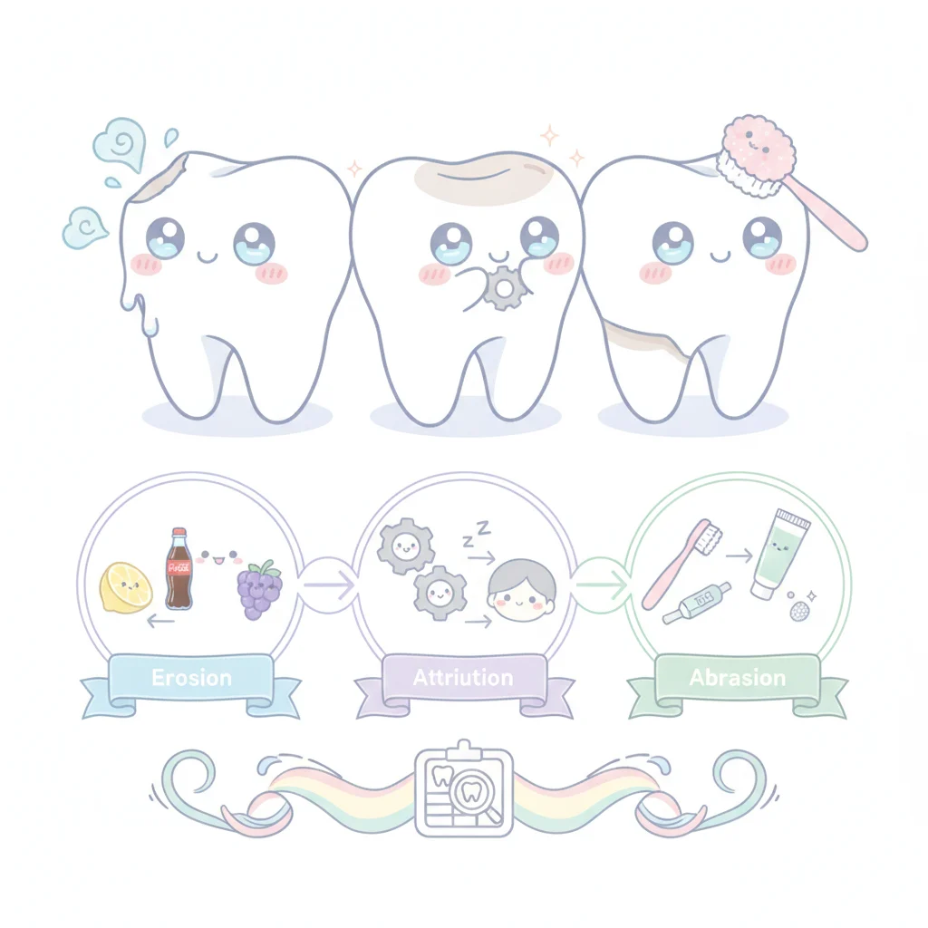
Erosion is the chemical dissolution of tooth structure by acids not produced by bacterial metabolism. This non-carious process leads to a characteristic smooth, scooped-out appearance, often with a dull, matte surface in early stages, progressing to dentin exposure and sensitivity. The primary culprits are extrinsic and intrinsic acids. Extrinsic sources include dietary acids from acidic beverages (soft drinks, fruit juices, sports drinks), acidic foods (citrus fruits, vinegar), and certain medications (aspirin, vitamin C chewables). Occupational exposure to acids, such as in battery factories or wine tasting, also contributes. Intrinsic sources are primarily gastric acids from conditions like gastroesophageal reflux disease (GERD), bulimia nervosa, or chronic vomiting due to other medical conditions. The pattern of erosion often provides clues: generalized erosion suggests dietary or environmental factors, while palatal erosion on maxillary anterior teeth is highly indicative of intrinsic acid exposure.
The mechanism of erosion involves the direct contact of acid with the enamel and dentin, leading to demineralization. Unlike caries, which involves bacterial plaque, erosion can occur on any tooth surface exposed to acid. The critical pH for enamel demineralization is approximately 5.5, but for dentin, it is higher, around 6.5, making dentin more susceptible to acid attack once exposed. Saliva plays a crucial protective role by buffering acids, clearing acidic substances, and providing calcium and phosphate ions for remineralization. Reduced salivary flow or altered salivary composition can significantly exacerbate erosive wear. Management of erosion focuses heavily on identifying and eliminating or mitigating the acid source. This might involve dietary counseling to reduce acidic food and drink intake, advising on proper timing of consumption, and avoiding brushing immediately after acid exposure when enamel is softened. For patients with GERD or bulimia, referral to a physician for medical management is paramount. Fluoride therapies, both topical and systemic, can increase enamel resistance to acid dissolution. Restorative interventions, such as bonding or crowns, are considered once the erosive process is controlled to prevent further loss and restore function and aesthetics.
Attrition: Wear from Tooth-to-Tooth Contact
Attrition is the physiological or pathological loss of tooth structure resulting from tooth-to-tooth contact during mastication or parafunctional habits like bruxism. This type of wear typically presents as flattened occlusal or incisal surfaces, often with sharp, well-defined facets that match opposing tooth surfaces. The wear can expose dentin, leading to sensitivity and a change in tooth morphology. While some degree of attrition is considered normal physiological wear over a lifetime, excessive attrition is usually pathological and indicates significant occlusal forces. Bruxism, whether awake or sleep-related, is a major contributor, involving repetitive clenching or grinding of teeth. Malocclusion, missing teeth, or poorly contoured restorations can also concentrate forces on specific teeth, accelerating attrition. The forces generated during bruxism can be considerably higher than those during normal chewing, leading to rapid and extensive wear.
The diagnosis of attrition relies on observing characteristic wear facets, often shiny and smooth, that correspond to opposing tooth surfaces. Patients may report jaw pain, headaches, muscle tenderness, or temporomandibular joint (TMJ) dysfunction. The management of attrition primarily involves protecting the teeth from excessive forces and addressing the underlying parafunctional habits. Occlusal splints or nightguards are commonly prescribed to absorb and distribute occlusal forces, preventing direct tooth-to-tooth contact during sleep bruxism. Behavioral therapies, stress management techniques, and biofeedback can help manage awake bruxism. Orthodontic treatment may be indicated to correct malocclusion and optimize occlusal relationships. Restorative treatments, such as crowns or onlays, are used to restore lost tooth structure, improve aesthetics, and re-establish proper occlusion once the causative factors are controlled. For a deeper dive into managing chronic conditions, clinicians might find our article on closing the gap in psoriasis and metabolic disease relevant for its multi-faceted approach to patient care.
Abrasion: External Mechanical Forces
Abrasion is the pathological loss of tooth structure due to mechanical forces from foreign objects habitually applied to the teeth. This is distinct from attrition, which involves tooth-to-tooth contact. The most common cause of abrasion is improper toothbrushing technique, particularly using a hard-bristled brush with excessive force or abrasive toothpastes. This typically results in V-shaped or U-shaped notches at the cervical margins of teeth, often more pronounced on the buccal surfaces. Other sources of abrasion include habits like chewing on pens, biting fingernails, using toothpicks excessively, or holding foreign objects between teeth (e.g., pipe stems, hairpins). Occupational abrasion can occur in individuals who hold tools or materials in their mouths. The pattern of abrasion is usually localized and directly related to the source of the mechanical force.
The mechanism involves the physical removal of tooth material by friction. The severity of abrasion depends on the intensity, frequency, and duration of the abrasive force, as well as the abrasiveness of the material. Enamel is relatively resistant, but dentin, once exposed, is much softer and more susceptible to abrasive wear. The diagnosis is typically straightforward, based on the characteristic appearance and location of the lesions, combined with a detailed patient history regarding oral hygiene habits and other oral behaviors. Management of abrasion focuses on eliminating the causative habit. This involves patient education on proper toothbrushing techniques, recommending soft-bristled brushes and low-abrasive toothpastes. For other habits, behavioral modification strategies are employed. Restorative treatments, such as composite resin restorations, are used to repair the lost tooth structure, reduce sensitivity, and improve aesthetics, but only after the abrasive habit has been addressed and controlled. Without addressing the underlying cause, any restoration will likely fail due to continued abrasive forces.
The Relationship of Wear Mechanisms
It is important to recognize that these three forms of tooth surface loss often do not occur in isolation. A patient may present with a combination of erosion, attrition, and abrasion, complicating diagnosis and treatment planning. For example, erosive lesions can soften enamel, making it more susceptible to attrition and abrasion. Similarly, areas of attrition or abrasion that expose dentin can then become more vulnerable to chemical erosion. This synergistic effect can accelerate tooth destruction. Clinicians must meticulously assess each patient to identify all contributing factors. A comprehensive treatment plan will then address each component, prioritizing the cessation of destructive habits and the control of underlying medical conditions. This integrated approach is essential for achieving long-term success and preventing further deterioration of the dentition. Understanding the relationship between these factors is key to effective patient education and compliance, which are vital for successful outcomes in managing tooth surface loss. The concept of small, single swaps for diet changes, for instance, can be adapted to oral hygiene habits, making behavioral modification more achievable for patients. This holistic view extends beyond the immediate dental presentation to consider broader patient health and lifestyle factors.
Diagnostic Challenges and Management Strategies
Distinguishing between these types of wear can be challenging, particularly when multiple factors are at play. A detailed history is indispensable, inquiring about dietary habits, medication use, medical conditions (especially those involving gastric reflux or eating disorders), and parafunctional habits. Clinical examination should include careful assessment of the location, shape, depth, and surface characteristics of the lesions. For instance, generalized cupping of cusps and incisal edges with intact enamel margins suggests erosion, while flat, shiny facets on opposing teeth point to attrition. V-shaped notches at the gingival margin are classic for abrasion. Sometimes, a combination of features will be present, requiring the clinician to determine the predominant factor. Diagnostic tools such as photographs, study models, and even occlusal analysis can aid in monitoring progression and guiding treatment. The HHS plan to accelerate clinical trial timelines highlights the broader push for efficient diagnostic and therapeutic pathways, a principle equally valuable in dental practice.
Management strategies are inherently tailored to the diagnosis. For erosion, the focus is on acid control, dietary modification, and fluoride application. For attrition, occlusal splints, behavioral therapy for bruxism, and occlusal adjustments are key. For abrasion, patient education on proper oral hygiene and cessation of harmful habits is paramount. Restorative treatments, ranging from simple composite fillings to full coverage crowns, are often necessary to repair lost tooth structure, restore function, and protect the pulp. But, restorations are only effective if the underlying cause of wear has been addressed. Placing a restoration without controlling the causative factor is akin to patching a leaky roof without fixing the hole; the problem will inevitably recur. Therefore, a phased approach, starting with etiological control and then moving to restorative care, is generally recommended. Regular follow-up and monitoring are essential to ensure the effectiveness of interventions and to detect any new or ongoing wear. The long-term success of managing tooth surface loss hinges on accurate diagnosis, comprehensive patient education, and a commitment to addressing the root causes, not just the symptoms.
The persistent challenge of tooth surface loss demands a more rigorous diagnostic approach from general practitioners. Simply noting 'wear' is insufficient; the specific etiology drives everything. Without distinguishing erosion from attrition or abrasion, clinicians risk prescribing ineffective or even harmful interventions, leaving patients vulnerable to continued dental destruction.
This precision in diagnosis is not merely academic; it directly impacts patient outcomes and resource allocation. Prescribing a nightguard for erosive wear, for example, is a waste of time and money, failing to address the underlying acid challenge. Conversely, dietary counseling for severe attrition due to bruxism misses the mark entirely.
For the industry, this highlights a need for better diagnostic tools and educational materials that emphasize etiological differentiation. Products designed to mitigate one form of wear may be useless for another. Patient education must also evolve, moving beyond generic advice to specific guidance tailored to their unique wear pattern.
The onus is on the clinician to perform the detective work. A detailed history and careful clinical examination are the most powerful tools available. Only then can a truly effective, preventive, and restorative plan be implemented, safeguarding patients' dentition for the long term.
- The Pivot Accurate diagnosis of the specific type of tooth surface loss (erosion, attrition, abrasion) is the critical first step.
- The Data No single numeric result applies, but the prevalence of tooth surface loss is increasing globally, affecting a significant portion of the adult population.
- The Action Tailor management plans to the identified etiology, focusing on prevention of further damage and conservative restorative approaches.
ART-2026-1387
·10/26
Drafted with AI assistance, reviewed and approved by the editorial team. This publication is intended for healthcare professionals, researchers, and life science industry professionals. Content is provided for informational and educational purposes only and does not constitute medical advice.

Digital health and patient experience are my beat: the apps, the wearables, the real-world evidence claims, and whether any of it changes outcomes. Sceptical by training and optimistic by instinct.
Cite This Article
Ward S, Voss M. Erosion, attrition, abrasion: why the cause changes the dental plan. The Life Science Feed. Published August 27, 2026. Updated October 1, 2026. Accessed October 11, 2026. https://thelifesciencefeed.com/dentistry/tooth-wear/practice/dental-wear-erosion-attrition-abrasion.
Editorial & AI Standards
All content is researched from peer-reviewed, open-access sources: published trial data, clinical guidelines, and regulatory filings. AI tools are used solely to structure and summarise that evidence; no AI-generated conclusions appear without editor verification against the primary source.
Every article is reviewed by a named editor before publication. Source citations are listed in the References section. This content does not represent the views of any pharmaceutical company, medical device manufacturer, or healthcare provider.
Licence & Rights
© 2026 The Life Science Feed. All rights reserved. Unless otherwise indicated, all content is the property of The Life Science Feed and may not be reproduced, distributed, or transmitted in any form or by any means without prior written permission.
Medical Disclaimer
The information provided on The Life Science Feed is for educational and informational purposes only. It is not intended as a substitute for professional medical advice, diagnosis, or treatment. Always seek the advice of your physician or other qualified healthcare provider regarding any medical condition or treatment decision. Never disregard professional medical advice or delay in seeking it because of something you have read on this website.
References
1. Warreth A, Abuhijleh E, Almaghribi MA, Mahwal G, Ashawish A. Tooth surface loss: A review of literature. Saudi Dent J. 2020;32(2):53-60. doi:10.1016/j.sdentj.2019.09.004
2. Dhaliwal G, Ouanounou A. Tooth surface loss: causes, management, and prevention. Quintessence Int. 2024;55(6):504-513. doi:10.3290/j.qi.b5223649
3. Abbott PV, Lin S. Tooth resorption-Part 2: A clinical classification. Dent Traumatol. 2022;38(4):267-285. doi:10.1111/edt.12762
4. Najeeb M, Islam S. Artificial intelligence (AI) in restorative dentistry: current trends and future prospects. BMC Oral Health. 2025;25(1):592. doi:10.1186/s12903-025-05989-1
5. Abbott PV. Prevention and management of external inflammatory resorption following trauma to teeth. Aust Dent J. 2016;61 Suppl 1:82-94. doi:10.1111/adj.12400
6. O'Hagan-Wong K, Enax J, Meyer F, Ganss B. The use of hydroxyapatite toothpaste to prevent dental caries. Odontology. 2022;110(2):223-230. doi:10.1007/s10266-021-00675-4