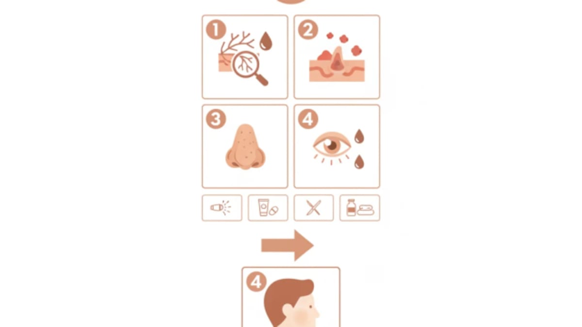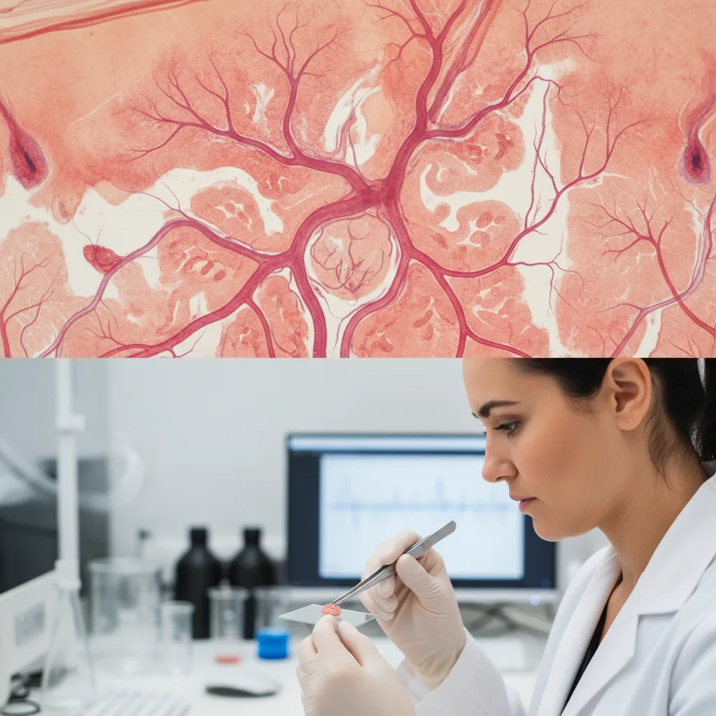

Rosacea, a chronic inflammatory skin condition, presents a significant challenge for clinicians, particularly when managing its hallmark symptom: persistent facial flushing. While erythema and papulopustular lesions often respond to established topical and systemic therapies, the episodic and often debilitating flushing frequently remains refractory. This therapeutic gap highlights a fundamental misunderstanding of rosacea's underlying pathophysiology, particularly the complex relationship between the nervous system and cutaneous vasculature.
Rosacea affects millions globally, manifesting as transient or persistent erythema, telangiectasias, papules, and pustules, predominantly on the central face. The condition carries a substantial burden on quality of life, with visible symptoms often leading to psychological distress and social anxiety. While the inflammatory lesions have seen advancements in treatment, the episodic flushing, often triggered by heat, stress, or certain foods, remains a persistent and frustrating symptom for many patients and their clinicians. This resistance to standard therapies suggests that the mechanisms driving flushing are distinct from those responsible for the inflammatory papules and pustules.
The traditional view of rosacea has largely focused on its inflammatory aspects, leading to the development of treatments that target immune responses and microbial factors. But the clinical reality of persistent flushing, which often precedes or occurs independently of other inflammatory signs, has pushed researchers to explore alternative pathophysiological pathways. This exploration has increasingly pointed towards a significant neurovascular component, where dysregulation of cutaneous blood vessels and their neural control plays a central role. Understanding this relationship is essential for developing more effective strategies for managing the most bothersome symptom for many patients.
The Neurovascular Hypothesis in Rosacea
The concept of rosacea as a purely inflammatory disorder has been challenged by observations that many patients experience prominent vascular symptoms, such as flushing and persistent erythema, without significant papulopustular involvement. This has led to a re-evaluation of the disease's origins, with a growing consensus that neurovascular dysregulation is a primary driver, particularly for the flushing phenotype. The skin's intricate vascular network is under constant modulation by the autonomic nervous system, neuropeptides, and local mediators, all of which appear to be perturbed in rosacea.
One key aspect of this hypothesis involves the transient receptor potential (TRP) channels, particularly TRPV1 and TRPA1. These ion channels are expressed on sensory neurons in the skin and act as polymodal sensors, responding to various stimuli such as heat, capsaicin, and environmental irritants. In rosacea, there is evidence of increased expression and sensitization of TRPV1 channels in cutaneous nerves and keratinocytes. This heightened sensitivity means that stimuli that would normally cause a mild vascular response in healthy individuals can trigger exaggerated flushing in rosacea patients. The activation of these channels leads to the release of neuropeptides, such as substance P and calcitonin gene-related peptide (CGRP), which are potent vasodilators. This mechanism explains why patients often report flushing in response to triggers like spicy foods or hot beverages, which directly activate these pathways.
Beyond TRP channels, other neural mechanisms contribute to flushing. The sympathetic nervous system, responsible for vasoconstriction and vasodilation, appears to be dysregulated. While sympathetic activation typically causes vasoconstriction, paradoxical vasodilation has been observed in rosacea patients in response to certain stimuli. This suggests an imbalance in adrenergic receptor function or an exaggerated response from cholinergic pathways. The involvement of the nervous system in conditions like excessive sweating also points to nerve dysfunction, a parallel that offers insights into rosacea's neurovascular underpinnings. For a deeper dive into related neurological mechanisms, consider our previous coverage on a new explanation for excessive sweating.
Vascular Dysfunction and Inflammatory Mediators
The vascular component of rosacea is not merely a passive recipient of neural signals; it actively contributes to the disease pathology. Patients with rosacea exhibit increased vascular reactivity and density of blood vessels, particularly telangiectasias. This increased vascularity is driven by angiogenic factors, such as vascular endothelial growth factor (VEGF), which are often elevated in rosacea skin. Chronic inflammation, even at a subclinical level, can perpetuate this angiogenic process, creating a vicious cycle where inflammation promotes vascular changes, which in turn exacerbate flushing and erythema.
Mast cells are another central player in this neurovascular dialogue. These immune cells are abundant in the skin and, when activated, release a plethora of vasoactive and pro-inflammatory mediators, including histamine, prostaglandins, and proteases. In rosacea, mast cells are often found in increased numbers and appear to be hyper-responsive, contributing significantly to both flushing and inflammation. The close proximity of mast cells to nerve endings and blood vessels facilitates a rapid and exaggerated response to various triggers, linking neural activation directly to vascular changes and inflammatory cascades. This intricate network of cellular and molecular interactions highlights the complexity of rosacea beyond a simple inflammatory process.
The role of cathelicidin, an antimicrobial peptide, has also been implicated in rosacea pathophysiology. In healthy skin, cathelicidin is processed into a form that has antimicrobial properties. But in rosacea, abnormal processing leads to an accumulation of a pro-inflammatory form of cathelicidin, LL-37. This aberrant LL-37 can induce inflammation, angiogenesis, and mast cell degranulation, directly contributing to both the inflammatory lesions and the vascular symptoms. This suggests a convergence of inflammatory and vascular pathways, where a single mediator can drive multiple aspects of the disease. The serotonin pathway also offers clues to rosacea's mechanism, further illustrating the complex neurochemical environment.
Why Standard Therapies Fall Short
Current standard therapies for rosacea primarily target the inflammatory components of the disease. Topical agents like metronidazole, azelaic acid, and ivermectin reduce papulopustular lesions by their anti-inflammatory and antimicrobial effects. Oral antibiotics, particularly subantimicrobial dose doxycycline, also work by inhibiting inflammatory pathways rather than their antibiotic action. These treatments are often effective for the inflammatory lesions but frequently provide limited relief for persistent flushing.
For the vascular symptoms, alpha-adrenergic agonists like brimonidine and oxymetazoline are used topically to induce vasoconstriction. These agents offer temporary relief by directly constricting cutaneous blood vessels, reducing erythema. But their effect is transient, and many patients experience rebound erythema or worsening flushing upon cessation, a phenomenon that highlights the underlying vascular dysregulation rather than resolving it. The fact that these direct vasoconstrictors only offer symptomatic relief, and often with caveats, highlights the need for therapies that address the root cause of the neurovascular dysfunction.
The limitations of existing treatments stem from their failure to adequately address the neural hypersensitivity and chronic vascular dysregulation that drive flushing. While they may dampen inflammation or temporarily constrict vessels, they do not reset the aberrant neurovascular signaling or reduce the underlying vascular reactivity. This is where the gap in treatment lies. Clinicians often find themselves managing symptoms rather than modifying disease progression for flushing, a challenge that can be particularly frustrating for patients. For comprehensive clinical guidance on managing various skin conditions, the Oxford Handbook of Medical Dermatology provides a step-by-step guide.
Future Directions: Targeting the Neurovascular Axis
The recognition of rosacea's neurovascular overlap opens new avenues for therapeutic development. Strategies that target TRP channels, neuropeptide release, or specific aspects of autonomic dysregulation could offer more sustained and effective relief for flushing. For example, antagonists of TRPV1 or CGRP receptors, currently being explored for other pain and migraine conditions, might find a role in rosacea. Modulating mast cell activity or inhibiting aberrant cathelicidin processing could also provide dual benefits by reducing both inflammation and vascular reactivity.
Non-pharmacological interventions, such as laser and light therapies, are already used for telangiectasias and persistent erythema. These modalities work by selectively targeting blood vessels, leading to their destruction or coagulation. While effective for visible vessels, their impact on the underlying neurovascular dysregulation and episodic flushing is less clear. Combining these physical therapies with novel pharmacological agents that address the neural component might offer a more comprehensive approach. The challenge remains in developing agents that can specifically modulate these complex pathways without causing systemic side effects.
The ongoing research into the precise mechanisms of neurovascular dysregulation in rosacea is of high importance for patients. Identifying specific biomarkers that correlate with flushing severity and treatment response would allow for more personalized therapeutic strategies. The heterogeneity of rosacea phenotypes also suggests that a one-size-fits-all approach is unlikely to succeed. Instead, tailoring treatments based on the predominant symptoms and underlying pathophysiological drivers, whether primarily inflammatory or neurovascular, will be essential for improving patient outcomes. This deeper understanding will hopefully lead to therapies that not only alleviate symptoms but also modify the disease course for those suffering from persistent and debilitating flushing.
The persistent challenge of rosacea flushing, despite advancements in treating inflammatory lesions, should prompt clinicians to think beyond a purely inflammatory paradigm. Standard anti-inflammatory or vasoconstrictive agents often provide only partial or temporary relief, leaving many patients with significant quality of life impairments. This suggests that the neurovascular component is not merely an adjunct but a central driver of this particular symptom.
When topical metronidazole or oral doxycycline fail to control flushing, it is a clear signal that the underlying neural hypersensitivity and vascular dysregulation are not being adequately addressed. Relying solely on temporary vasoconstrictors like brimonidine, with its known rebound effect, is a symptomatic bandage rather than a therapeutic solution. We need to acknowledge that the disease mechanism for flushing is distinct and requires a different approach.
The industry's focus on anti-inflammatory pathways has been productive for papulopustular rosacea, but the market for effective, sustained flushing treatments remains largely unmet. Future drug development must pivot towards modulating TRP channels, neuropeptide release, or mast cell activity to truly impact this debilitating symptom. Until then, clinicians must manage expectations and consider combination strategies that acknowledge the complex relationship of neural and vascular factors.
- The Pivot Rosacea flushing is increasingly understood as a neurovascular disorder, not solely an inflammatory one, explaining its resistance to anti-inflammatory treatments.
- The Data Current treatments primarily target inflammatory pathways, leaving the neural and vascular dysregulation largely unaddressed.
- The Action Clinicians should consider the neurovascular component of rosacea when standard anti-inflammatory or vasoconstrictive therapies prove insufficient for flushing.
ART-2026-1234
·10/26
Drafted with AI assistance, reviewed and approved by the editorial team. This publication is intended for healthcare professionals, researchers, and life science industry professionals. Content is provided for informational and educational purposes only and does not constitute medical advice.

I cover women's health, reproductive medicine, and the persistent gaps in how conditions that primarily affect women get studied and funded. The evidence base is thinner than it should be. I write about why.
Cite This Article
Mitchell S, Voss M. Why rosacea flushing resists standard therapy: a neurovascular disconnect. The Life Science Feed. Published August 27, 2026. Updated October 6, 2026. Accessed October 12, 2026. https://thelifesciencefeed.com/dermatology/rosacea/insights/rosacea-flushing-neurovascular-overlap-therapy-resistance.
Editorial & AI Standards
All content is researched from peer-reviewed, open-access sources: published trial data, clinical guidelines, and regulatory filings. AI tools are used solely to structure and summarise that evidence; no AI-generated conclusions appear without editor verification against the primary source.
Every article is reviewed by a named editor before publication. Source citations are listed in the References section. This content does not represent the views of any pharmaceutical company, medical device manufacturer, or healthcare provider.
Licence & Rights
© 2026 The Life Science Feed. All rights reserved. Unless otherwise indicated, all content is the property of The Life Science Feed and may not be reproduced, distributed, or transmitted in any form or by any means without prior written permission.
Medical Disclaimer
The information provided on The Life Science Feed is for educational and informational purposes only. It is not intended as a substitute for professional medical advice, diagnosis, or treatment. Always seek the advice of your physician or other qualified healthcare provider regarding any medical condition or treatment decision. Never disregard professional medical advice or delay in seeking it because of something you have read on this website.
References
1. Sharma A, Kroumpouzos G, Kassir M, et al. Rosacea management: A comprehensive review. J Cosmet Dermatol. 2022;21(5):1895-1904. doi:10.1111/jocd.14816
2. Mohamed-Noriega K, Loya-Garcia D, Vera-Duarte GR, et al. Ocular Rosacea: An Updated Review. Cornea. 2025;44(4):525-537. doi:10.1097/ICO.0000000000003785
3. Ivanic MG, Oulee A, Norden A, Javadi SS, Gold MH, Wu JJ. Neurogenic Rosacea Treatment: A Literature Review. J Drugs Dermatol. 2023;22(6):566-575. doi:10.36849/JDD.7181
4. Avraham S, Khaslavsky S, Kashetsky N, et al. Treatment of ocular rosacea: a systematic review. J Dtsch Dermatol Ges. 2024;22(2):167-174. doi:10.1111/ddg.15290
5. van Zuuren EJ, Arents BWM, van der Linden MMD, Vermeulen S, Fedorowicz Z, Tan J. Rosacea: New Concepts in Classification and Treatment. Am J Clin Dermatol. 2021;22(4):457-465. doi:10.1007/s40257-021-00595-7
6. Gether L, Overgaard LK, Egeberg A, Thyssen JP. Incidence and prevalence of rosacea: a systematic review and meta-analysis. Br J Dermatol. 2018;179(2):282-289. doi:10.1111/bjd.16481