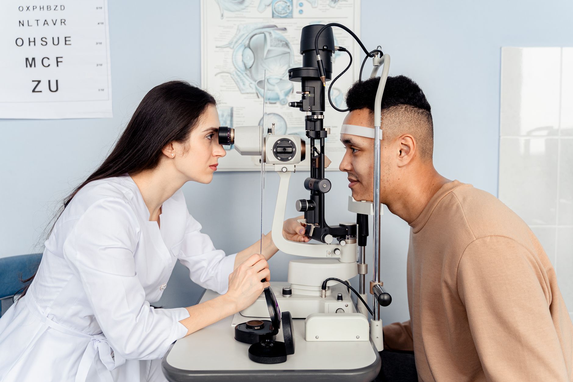

Glaucoma management traditionally focuses on intraocular pressure reduction, but a growing body of evidence highlights a critical, often neglected, comorbidity: ocular surface disease (OSD). This condition, encompassing dry eye and other surface pathologies, significantly complicates glaucoma care, affecting patient comfort, treatment adherence, and overall quality of life. Clinicians must integrate OSD evaluation into routine glaucoma practice to improve outcomes.1
Glaucoma, a leading cause of irreversible blindness, demands lifelong management, primarily through topical hypotensive medications. These drops, while effective at lowering intraocular pressure (IOP), often come with a significant burden of ocular surface disease (OSD), including dry eye, blepharitis, and conjunctival inflammation. For too long, clinicians have considered OSD a mere nuisance, a predictable side effect of chronic topical therapy. But new insights challenge this narrow view, positioning OSD as an essential, integrated component of glaucoma care.1
Dr. Morten Kolko, in his comprehensive review published in J Glaucoma, argues for a fundamental shift in how the ophthalmic community approaches OSD in glaucoma patients.1 Kolko emphasizes that OSD is not solely an iatrogenic consequence but often predates glaucoma diagnosis or exacerbates existing conditions, creating a vicious cycle that compromises both patient well-being and treatment efficacy. This perspective demands a proactive, rather than reactive, approach to ocular surface health.1
The Pathophysiological Overlap
The connection between glaucoma and OSD runs deeper than simple irritation from eye drops. Kolko details several shared pathophysiological mechanisms. Chronic inflammation, a hallmark of OSD, also plays a role in glaucomatous optic neuropathy. Inflammatory mediators, such as cytokines and chemokines, found in the tears and ocular surface of OSD patients, can contribute to neuroinflammation and retinal ganglion cell damage. This suggests a bidirectional relationship where inflammation on the surface may influence the progression of glaucoma itself.1
Oxidative stress is another common denominator. Both glaucoma and OSD involve increased oxidative damage to ocular tissues. In OSD, this stress contributes to goblet cell loss, epithelial damage, and tear film instability. In glaucoma, oxidative stress is implicated in optic nerve head damage. The cumulative effect of these stressors on the ocular surface and the optic nerve creates a more vulnerable eye, potentially accelerating disease progression and worsening symptoms.1
Neurosensory abnormalities also link the two conditions. Patients with OSD often exhibit corneal nerve damage and reduced corneal sensitivity, leading to diminished reflex tearing and persistent discomfort. Glaucoma patients, particularly those on long-term topical therapy, can also develop corneal neuropathy. This impaired corneal sensation can mask the severity of OSD symptoms, leading to underdiagnosis and undertreatment, even as the ocular surface continues to deteriorate.1
The Impact of Topical Glaucoma Medications
While OSD is not exclusively treatment-induced, topical glaucoma medications undeniably exacerbate or initiate it in many patients. Preservatives, particularly benzalkonium chloride (BAK), are major culprits. BAK causes direct toxicity to corneal and conjunctival epithelial cells, goblet cells, and meibomian glands. It disrupts the tear film, increases inflammatory markers, and can lead to permanent changes in the ocular surface architecture.1
Even preservative-free formulations, while generally better tolerated, are not entirely benign. The active pharmaceutical ingredients themselves can contribute to OSD. Beta-blockers can reduce aqueous tear production, prostaglandin analogues can induce conjunctival hyperemia and meibomian gland dysfunction, and carbonic anhydrase inhibitors can cause local irritation. The sheer volume and frequency of drops, especially in patients requiring multiple medications, further stress the ocular surface.1
The cumulative effect of these factors leads to a high prevalence of OSD in glaucoma patients, ranging from 40% to 90% depending on the diagnostic criteria used.1 This is significantly higher than the general population prevalence of dry eye, which typically hovers around 10-20%. The discrepancy highlights the unique challenges faced by glaucoma patients and the need for targeted interventions.1
Diagnosis: Beyond the Obvious
Kolko stresses that OSD diagnosis in glaucoma patients requires a systematic approach, moving beyond patient-reported symptoms alone. Many glaucoma patients, especially older individuals, may have reduced corneal sensitivity or may attribute their discomfort to aging or their primary glaucoma. This leads to significant underreporting of symptoms.1
Validated questionnaires, such as the Ocular Surface Disease Index (OSDI) or the Dry Eye Questionnaire 5 (DEQ-5), are essential screening tools. These instruments quantify symptom severity and frequency, providing a baseline and allowing for monitoring of treatment response. A score above a certain threshold, for example, OSDI > 22, indicates moderate to severe OSD.1
Objective clinical signs are equally important. Tear film break-up time (TBUT) measures tear film stability; a TBUT of <10 seconds is indicative of dry eye. Fluorescein staining of the cornea and conjunctiva can reveal epithelial damage, with punctate keratitis being a common finding. Lissamine green or rose bengal staining can highlight conjunctival damage, particularly in the interpalpebral region. Schirmer's test, while less specific, can assess aqueous tear production, with values <5 mm in 5 minutes suggesting aqueous deficiency.1
Meibography, which images the meibomian glands, can detect meibomian gland dysfunction (MGD), a frequent comorbidity. MGD contributes to evaporative dry eye and is often overlooked. Inflammatory markers, such as matrix metalloproteinase-9 (MMP-9) detected via point-of-care tests, can also provide objective evidence of ocular surface inflammation. Integrating these diagnostic modalities allows for a comprehensive assessment, ensuring that OSD is not missed.1
Management Strategies: An Integrated Approach
Effective OSD management in glaucoma patients requires a multi-pronged strategy. The first step involves optimizing glaucoma therapy itself. Where possible, clinicians should consider preservative-free glaucoma medications, especially for patients on multiple drops or those with pre-existing OSD. Switching from BAK-preserved drops to non-BAK preserved or preservative-free options can significantly improve OSD symptoms and signs.1
Selective laser trabeculoplasty (SLT) or minimally invasive glaucoma surgery (MIGS) can reduce or eliminate the need for topical medications, thereby alleviating OSD. For patients whose IOP is well-controlled with drops but suffer severe OSD, these surgical options become particularly attractive. The decision to pursue surgical intervention for OSD relief, however, must be balanced against the risks and benefits of the procedure and the patient's glaucoma status.1
Beyond glaucoma medication adjustments, direct OSD treatment is essential for patient comfort and treatment adherence. Artificial tears, preferably preservative-free, are the first-line therapy for mild to moderate dry eye. These drops provide lubrication and help stabilize the tear film. For more severe cases, topical anti-inflammatory agents, such as cyclosporine or lifitegrast, can reduce ocular surface inflammation. These medications target the underlying inflammatory processes contributing to OSD.1
Lid hygiene, warm compresses, and oral omega-3 fatty acid supplementation can address MGD and blepharitis. Punctal plugs can conserve natural tears in patients with aqueous deficient dry eye. Autologous serum tears, derived from the patient's own blood, offer a more advanced option for severe, refractory OSD, providing growth factors and vitamins that promote ocular surface healing.1
The Catch: Adherence and Quality of Life
The implications of untreated OSD extend beyond patient discomfort. Poor ocular surface health directly impacts adherence to glaucoma medication. Patients experiencing significant burning, stinging, or foreign body sensation are less likely to take their drops consistently. This non-adherence, in turn, leads to uncontrolled IOP and an increased risk of glaucoma progression.1
Quality of life is also severely affected. Chronic ocular discomfort can lead to anxiety, depression, and reduced ability to perform daily activities, such as reading or driving. The burden of managing two chronic eye conditions can be overwhelming for patients, highlighting the need for a holistic approach that considers both IOP control and ocular surface health.1
"Ignoring ocular surface disease in glaucoma patients is akin to treating hypertension without considering the patient's diet. It's a fundamental oversight that compromises long-term success."Morten Kolko, J Glaucoma 2026
The open-label nature of many OSD studies is an obvious caveat, as subjective symptom improvement can be influenced by patient expectation. Still, objective measures like TBUT and staining scores provide a more robust assessment. The challenge remains in conducting large-scale, randomized controlled trials specifically designed to evaluate the impact of integrated OSD management on glaucoma progression and long-term outcomes.1
Kolko's review serves as a powerful call to action for clinicians. Integrating OSD evaluation and management into routine glaucoma care is not an optional add-on but an essential component for improving patient comfort, medication adherence, and ultimately, preserving vision. The Oxford Handbook of Ophthalmology provides a concise guide to ophthalmic practice, including detailed sections on both glaucoma and ocular surface disease, which can aid clinicians in this integrated approach. The next step for the field involves developing clear, evidence-based guidelines for OSD screening and treatment pathways tailored specifically for glaucoma patients.
The message from Kolko is unambiguous: ocular surface disease is not a secondary concern in glaucoma management. Clinicians who continue to view OSD as a mere side effect of topical drops are missing a significant piece of the patient's clinical picture, one that directly impacts adherence and, by extension, IOP control. This demands a proactive shift from reactive symptom management to routine, systematic screening.
For patients, the implications are profound. Chronic discomfort from OSD can be debilitating, leading to a vicious cycle of non-adherence to sight-saving glaucoma medications. Improving ocular surface health can dramatically enhance their quality of life and ensure they remain on their prescribed regimens, ultimately preserving vision more effectively than focusing solely on IOP numbers.
The pharmaceutical industry also has a role to play. While preservative-free options are becoming more common, the development of novel glaucoma therapies with minimal ocular surface impact remains a priority. Better integration of OSD assessment into clinical trial endpoints for new glaucoma drugs would provide a more complete understanding of their real-world tolerability.
The onus is on individual practitioners to adopt this integrated approach. A quick OSDI questionnaire or a rapid TBUT assessment during a routine glaucoma follow-up could uncover significant, treatable issues that are currently being overlooked. This small change in practice could yield substantial benefits for patients struggling with the dual burden of glaucoma and ocular surface disease.
- The Pivot Ocular surface disease is not merely a side effect of glaucoma treatment but an intrinsic, pathophysiologically linked component of the disease itself.
- The Data OSD prevalence in glaucoma patients ranges from 40% to 90%, with symptoms often underreported and undertreated.1
- The Action Routinely screen all glaucoma patients for OSD using validated questionnaires and objective clinical signs to enable early intervention and improve treatment tolerability.
ART-2026-1112
·10/26
Drafted with AI assistance, reviewed and approved by the editorial team. This publication is intended for healthcare professionals, researchers, and life science industry professionals. Content is provided for informational and educational purposes only and does not constitute medical advice.

Science writer covering the frontier between basic research and clinical practice. I am interested in the moment a mechanism becomes a therapy, and everything that can go wrong in between.
Cite This Article
Aldrich M, Voss M. Ocular surface disease: the overlooked comorbidity complicating glaucoma management. The Life Science Feed. Published August 20, 2026. Updated October 3, 2026. Accessed October 4, 2026. https://thelifesciencefeed.com/ophthalmology/glaucoma/practice/ocular-surface-disease-the-overlooked-comorbidity-complicating-glaucoma-management.
Editorial & AI Standards
All content is researched from peer-reviewed, open-access sources: published trial data, clinical guidelines, and regulatory filings. AI tools are used solely to structure and summarise that evidence; no AI-generated conclusions appear without editor verification against the primary source.
Every article is reviewed by a named editor before publication. Source citations are listed in the References section. This content does not represent the views of any pharmaceutical company, medical device manufacturer, or healthcare provider.
Licence & Rights
© 2026 The Life Science Feed. All rights reserved. Unless otherwise indicated, all content is the property of The Life Science Feed and may not be reproduced, distributed, or transmitted in any form or by any means without prior written permission.
Medical Disclaimer
The information provided on The Life Science Feed is for educational and informational purposes only. It is not intended as a substitute for professional medical advice, diagnosis, or treatment. Always seek the advice of your physician or other qualified healthcare provider regarding any medical condition or treatment decision. Never disregard professional medical advice or delay in seeking it because of something you have read on this website.
References
1. Kolko M. Rethinking Dry Eye and Ocular Surface Disease in Glaucoma: Pathophysiology, Diagnosis, and Management. J Glaucoma 2026.







