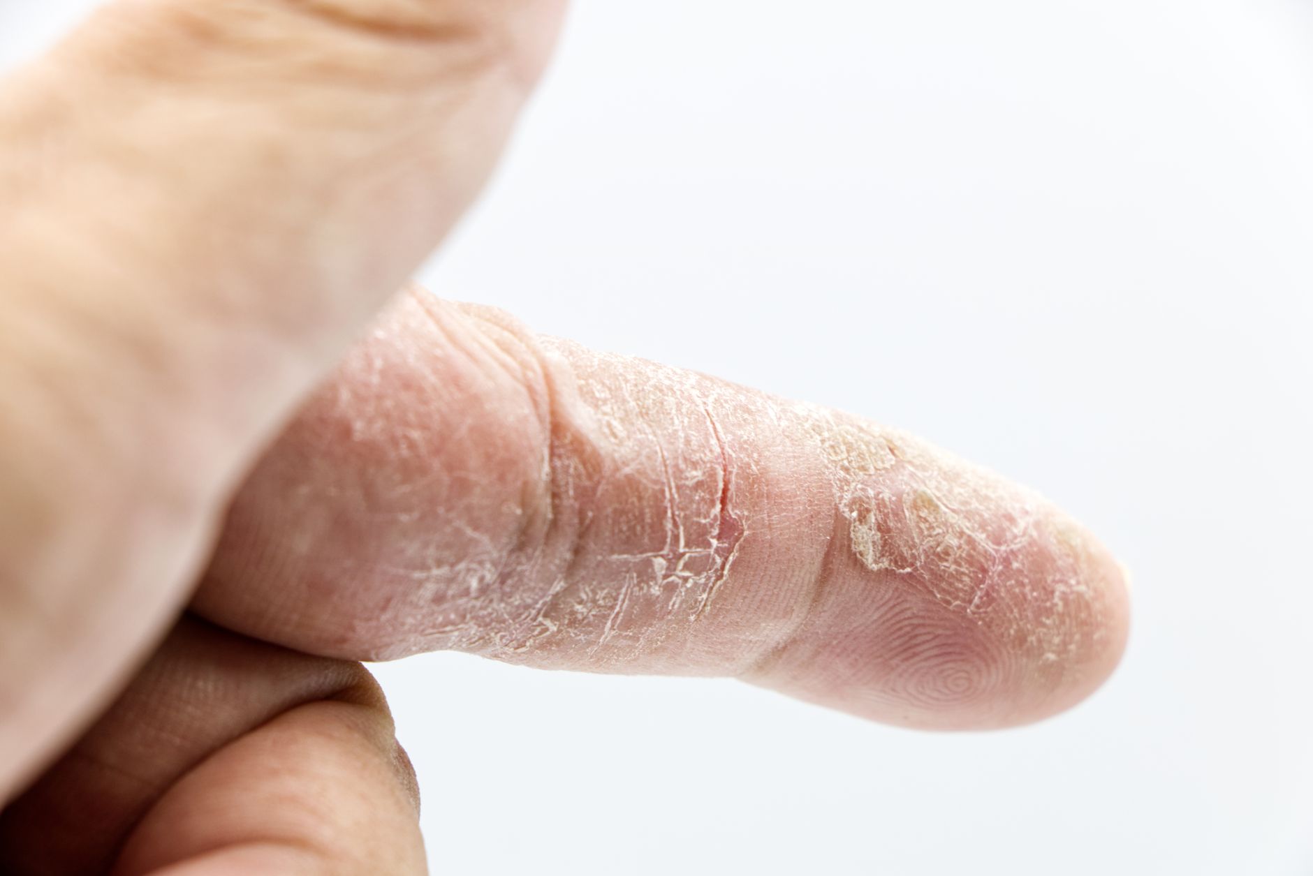

Harlequin ichthyosis presents as one of the most severe forms of congenital ichthyosis, a condition immediately apparent at birth due to profound cutaneous abnormalities. This rare genetic disorder transforms the skin into thick, plate-like scales, often leading to life-threatening complications in neonates. Understanding its precise genetic and pathophysiological underpinnings is critical for early diagnosis and management, offering a clearer path for clinicians navigating this complex presentation. The Medscape article on Harlequin Ichthyosis provides a comprehensive overview of its background, etiology, and epidemiology.
Harlequin ichthyosis (HI) represents the most severe manifestation within the spectrum of congenital ichthyoses, a group of genetic disorders characterized by generalized scaling of the skin. Affected neonates are born encased in thick, hyperkeratotic plates separated by deep fissures, giving the skin a distinctive, armor-like appearance. This profound disruption of the epidermal barrier leads to significant fluid loss, electrolyte imbalance, thermoregulatory dysfunction, and increased susceptibility to infection, posing an immediate and severe threat to life. Historically, HI carried an exceptionally grim prognosis, with most infants succumbing within days or weeks of birth. Advances in neonatal intensive care, however, have shifted this outlook, allowing a growing number of patients to survive the critical neonatal period and transition into chronic management.1
The clinical presentation of HI is striking and unmistakable at birth. The characteristic thick, yellowish, diamond-shaped scales cover the entire body, often pulling the facial features taut. This tension leads to severe ectropion (eversion of the eyelids), eclabium (eversion of the lips), and flattened ears and nose. The hands and feet may be fixed in flexion contractures, and digits can be hypoplastic or even amputated due to constricting bands of skin. Respiratory distress is common, resulting from restricted chest wall movement and aspiration risk. These severe physical manifestations are a direct consequence of the underlying genetic defect, which fundamentally compromises the skin's ability to form an effective barrier.1
The Genetic Blueprint of a Compromised Barrier
Harlequin ichthyosis is an autosomal recessive disorder, meaning an individual must inherit two copies of the mutated gene, one from each parent, to develop the condition. The causative gene is ABCA12, located on chromosome 2q35. This gene encodes for an ATP-binding cassette (ABC) transporter protein, specifically ABCA12, which plays a critical role in lipid transport within keratinocytes. The ABCA12 protein is essential for the normal development of the lamellar granules, organelles within epidermal cells that package and secrete lipids into the extracellular space. These lipids are crucial for forming the lipid bilayers that constitute the skin's impermeable barrier.1
Mutations in ABCA12 lead to a dysfunctional or absent ABCA12 protein. Without a properly functioning ABCA12 transporter, keratinocytes cannot adequately process and transport lipids into lamellar granules. This results in a severe deficiency of barrier lipids, particularly ceramides, in the stratum corneum. The consequence is a profoundly impaired epidermal barrier, unable to prevent excessive transepidermal water loss (TEWL) or protect against external pathogens. The hyperkeratosis and scaling observed in HI are a compensatory response to this defective barrier, as the skin attempts to overproduce keratinocytes to form some semblance of protection.1
Epidemiology and Incidence
Harlequin ichthyosis is exceedingly rare, with an estimated incidence ranging from 1 in 300,000 to 1 in 1,000,000 live births. Due to its autosomal recessive inheritance pattern, the disorder affects males and females equally. While no specific ethnic predilection has been definitively established, cases have been reported across diverse populations worldwide. The rarity of the condition means that many clinicians, even those in specialized fields, may encounter only one or two cases in their entire careers, if any. This limited exposure underscores the importance of readily accessible, comprehensive information for prompt diagnosis and appropriate management.1
The prevalence of carrier status for ABCA12 mutations in the general population is unknown, but given the rarity of the disease, it is presumed to be low. Consanguinity increases the risk of affected offspring, as it raises the probability of two carriers mating and passing on the same recessive mutation. Genetic counseling is therefore a critical component of care for families with a history of HI, allowing for informed reproductive choices and prenatal diagnosis in subsequent pregnancies.1
Diagnosis and Differential Considerations
Diagnosis of Harlequin ichthyosis is typically made clinically at birth due to the unmistakable phenotype. Prenatal diagnosis is possible through genetic testing of amniotic fluid cells or chorionic villus samples, particularly in families with a known history of the disorder or identified ABCA12 mutations. Ultrasonography may reveal suggestive features such as polyhydramnios, fetal growth restriction, or abnormal facial features, but these are non-specific. Postnatally, a skin biopsy can confirm the diagnosis by demonstrating characteristic histopathological features, including marked hyperkeratosis, parakeratosis, and abnormal lamellar granule formation. Genetic testing for ABCA12 mutations provides definitive confirmation.1
The differential diagnosis for severe congenital ichthyosis includes other forms of ichthyosis, such as lamellar ichthyosis and congenital ichthyosiform erythroderma. However, the extreme severity and characteristic plate-like scales of HI usually distinguish it from these milder presentations. Collodion baby, a phenotype where an infant is born encased in a tight, shiny membrane, can be a precursor to several types of ichthyosis, including HI, but the subsequent development of the thick, fissured scales of HI is unique. A thorough clinical examination combined with genetic analysis is essential to avoid misdiagnosis and ensure appropriate management.1
Pathophysiology Beyond the Barrier
While the primary defect in HI lies in the epidermal barrier, the systemic consequences extend far beyond the skin. The constant battle against fluid loss and infection places immense metabolic strain on affected infants. Dehydration and electrolyte imbalances, particularly hypernatremia, are common and require aggressive intravenous fluid resuscitation. The deep fissures in the skin serve as portals of entry for bacteria, leading to sepsis, a frequent cause of mortality in the neonatal period. Thermoregulation is also severely impaired; the rigid skin plates prevent normal sweating, leading to hyperthermia, while the extensive skin damage also causes significant heat loss.1
Respiratory compromise is another major concern. The tight, constricting skin around the chest wall can restrict lung expansion, leading to respiratory distress. Ectropion and eclabium can interfere with feeding and increase the risk of aspiration pneumonia. The constant tension on the facial skin can also lead to corneal damage and visual impairment if not managed meticulously. These multifaceted challenges necessitate a multidisciplinary approach to care, involving neonatologists, dermatologists, ophthalmologists, and geneticists. The Oxford Handbook of Paediatrics offers a concise guide to managing such complex neonatal conditions.
Management Strategies and Prognosis
Management of Harlequin ichthyosis in the neonatal period is primarily supportive and intensive. Infants require immediate admission to a neonatal intensive care unit (NICU). Aggressive hydration with intravenous fluids is crucial to combat dehydration and electrolyte disturbances. Maintaining a humidified environment helps to reduce transepidermal water loss. Topical emollients, often petroleum-based, are applied frequently and generously to soften the scales and improve skin pliability. Systemic retinoids, such as isotretinoin or acitretin, have shown promise in reducing hyperkeratosis and improving skin barrier function, though their use in neonates requires careful consideration due to potential side effects.1
Infection prevention is paramount. Broad-spectrum antibiotics are often initiated empirically and adjusted based on culture results. Ophthalmic lubricants and protective measures are necessary to prevent corneal damage from ectropion. Nutritional support, often via nasogastric tube, is essential to meet the high metabolic demands. As patients survive the neonatal period, long-term management focuses on chronic skin care, prevention of complications, and addressing developmental needs. Physical therapy may be required to manage contractures. Genetic counseling remains vital for families, providing information on recurrence risk and reproductive options. While the prognosis has improved, HI remains a severe, lifelong condition requiring continuous, specialized care.1
The improved survival rates for Harlequin ichthyosis neonates mean GPs and specialists will increasingly encounter these patients beyond the immediate critical care phase. This shift necessitates a broader understanding of the long-term dermatological, developmental, and psychosocial challenges these individuals face. The initial shock of the diagnosis for parents requires sensitive and informed counseling, extending beyond the immediate medical crisis to encompass genetic implications and chronic care planning.
For clinicians in primary care, recognizing the signs of infection, managing chronic skin dryness, and coordinating multidisciplinary follow-up will become standard practice. The ongoing need for specialized emollients and potentially systemic retinoids means close collaboration with dermatologists is essential. These patients often require frequent monitoring for complications like contractures, visual impairment, and growth delays, demanding a proactive and integrated approach to care.
The rarity of Harlequin ichthyosis means that diagnostic delays can occur, particularly in settings without immediate access to specialized neonatal or dermatological expertise. Educating healthcare providers on the distinctive clinical features and the importance of early intervention can significantly impact initial outcomes. Furthermore, the psychological burden on families is immense, and connecting them with support groups and genetic counseling services is a crucial, often overlooked, aspect of comprehensive care.
- The Pivot Harlequin ichthyosis, once almost universally fatal, now sees improved survival rates due to advances in neonatal intensive care and targeted dermatological management.
- The Data The disorder is caused by autosomal recessive mutations in the ABCA12 gene, located on chromosome 2q35, which encodes an ATP-binding cassette transporter.
- The Action Early recognition, aggressive supportive care, and genetic counseling for affected families are paramount for improving outcomes and informing reproductive decisions.
ART-2026-970
·10/26
Drafted with AI assistance and checked by automated style rules. Not individually read by an editor. This publication is intended for healthcare professionals, researchers, and life science industry professionals. Content is provided for informational and educational purposes only and does not constitute medical advice.

I write about AI in healthcare: the validation studies, the deployment failures, and the regulatory questions without answers yet. Based in San Francisco, close to where the technology is built.
Cite This Article
Chen L, Voss M. Harlequin ichthyosis: understanding the genetic basis of a rare skin disorder. The Life Science Feed. Published July 26, 2026. Updated October 6, 2026. Accessed October 24, 2026. https://thelifesciencefeed.com/rare-diseases/cystic-fibrosis/insights/harlequin-ichthyosis-understanding-the-genetic-basis-of-a-rare-skin-disorder.
Editorial & AI Standards
Content is drafted with AI tools from peer-reviewed, open-access sources such as published trial data, clinical guidelines, and regulatory filings. This article is checked by automated style rules but is not individually read by an editor, so check the cited source before relying on a figure.
Source citations are listed in the References section. This content does not represent the views of any pharmaceutical company, medical device manufacturer, or healthcare provider.
Licence & Rights
© 2026 The Life Science Feed. All rights reserved. Unless otherwise indicated, all content is the property of The Life Science Feed and may not be reproduced, distributed, or transmitted in any form or by any means without prior written permission.
Medical Disclaimer
The information provided on The Life Science Feed is for educational and informational purposes only. It is not intended as a substitute for professional medical advice, diagnosis, or treatment. Always seek the advice of your physician or other qualified healthcare provider regarding any medical condition or treatment decision. Never disregard professional medical advice or delay in seeking it because of something you have read on this website.
References
1. Harlequin Ichthyosis: Background, Etiology, Epidemiology. Medscape. Updated October 27,
2023. Accessed January 1,
2024. https://www.medscape.com/answers/1111604-106456/what-is-the-background-of-harlequin-ichthyosis