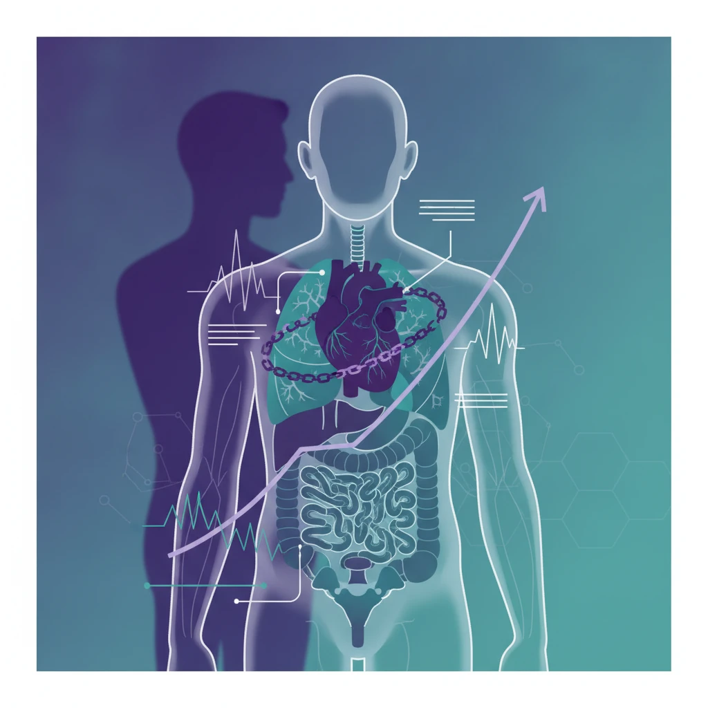

Duchenne muscular dystrophy (DMD) is a progressive, X-linked genetic disorder primarily affecting skeletal muscle, but its systemic reach extends critically to the heart and lungs. While muscle weakness defines the early stages, cardiomyopathy and respiratory failure emerge as the leading causes of morbidity and mortality in later disease. Effective management hinges on rigorous, scheduled surveillance of these vital systems.
The natural history of DMD has shifted dramatically over the past few decades, not due to a single therapeutic breakthrough, but largely through the consistent application of multidisciplinary care, particularly in cardiac and respiratory management. This proactive approach has transformed DMD from a childhood disease with a grim prognosis to a condition where many patients now live into adulthood, underscoring the profound impact of timely intervention.
Duchenne muscular dystrophy, caused by mutations in the dystrophin gene, leads to the absence or dysfunction of the dystrophin protein, which is essential for muscle cell integrity. Without functional dystrophin, muscle fibers are highly susceptible to damage during contraction, leading to cycles of degeneration and regeneration that eventually deplete the muscle stem cell pool and result in fibrosis and fat replacement. This process affects all striated muscles, including the diaphragm, intercostal muscles, and the myocardium, leading to predictable patterns of cardiac and respiratory decline.
The progressive nature of DMD means that while ambulation may be lost in early adolescence, the insidious onset of cardiac and respiratory complications often begins much earlier, frequently before symptoms become overt. This silent progression necessitates a structured approach to surveillance, designed to catch these complications at their earliest, most treatable stages. The goal is not merely to react to symptoms, but to anticipate and mitigate the inevitable decline, thereby preserving function and extending life.
The Cardiac Imperative: Early Detection of Cardiomyopathy
Duchenne cardiomyopathy is a distinct entity, typically presenting as a dilated cardiomyopathy, though left ventricular non-compaction and arrhythmias can also occur. It is often asymptomatic for prolonged periods, even as significant myocardial damage accumulates. The standard of care mandates regular cardiac evaluations, beginning from diagnosis or by age 10 at the latest, whichever comes first. These evaluations typically involve echocardiography and electrocardiography (ECG).
Echocardiography is the cornerstone of cardiac surveillance, providing critical information on left ventricular ejection fraction (LVEF), chamber dimensions, and wall motion abnormalities. Guidelines recommend annual echocardiograms once cardiomyopathy is suspected or diagnosed, but earlier and more frequent screening may be warranted in patients with specific dystrophin mutations or those on certain therapies. The Braunwald's Heart Disease textbook provides extensive detail on the pathophysiology and management of various cardiomyopathies, including those seen in muscular dystrophies.
Cardiac magnetic resonance imaging (CMR) offers a more sensitive assessment of myocardial fibrosis, a hallmark of Duchenne cardiomyopathy, even before changes in LVEF are apparent. CMR can detect subtle myocardial damage and quantify fibrosis, which may predict future cardiac dysfunction. While not universally recommended for annual screening due to accessibility and cost, CMR is increasingly used as a baseline assessment and for clarifying equivocal echocardiographic findings. Early identification of fibrosis can prompt earlier initiation of cardioprotective therapies.
Electrocardiography is also a vital component, primarily to detect arrhythmias and conduction abnormalities. Sinus tachycardia, short PR interval, and tall R waves in V1 are common findings in DMD patients. While these may not always require immediate intervention, they can signal underlying myocardial stress or fibrosis. Regular ECGs, typically annually, help monitor for changes that might necessitate further investigation or antiarrhythmic therapy.
Pharmacological interventions for Duchenne cardiomyopathy typically involve angiotensin-converting enzyme (ACE) inhibitors or angiotensin receptor blockers (ARBs), often initiated even in the presence of preserved LVEF, particularly if myocardial fibrosis is detected. Beta-blockers are added once LVEF begins to decline. These therapies aim to reduce cardiac workload, prevent ventricular remodeling, and slow the progression of heart failure. The timing of initiation is crucial for patient survival; starting these agents before overt symptoms or significant LVEF reduction can significantly delay the onset of symptomatic heart failure and improve long-term outcomes. This proactive approach mirrors the strategy for cardiac amyloidosis diagnoses, where early detection is paramount.
Navigating Respiratory Decline: The Pulmonary Challenge
Respiratory muscle weakness in DMD progresses relentlessly, affecting both the diaphragm and intercostal muscles. This leads to restrictive lung disease, impaired cough efficacy, and ultimately, chronic respiratory insufficiency. Like cardiomyopathy, respiratory compromise often develops insidiously, with nocturnal hypoventilation preceding daytime symptoms. This makes regular, objective assessment indispensable.
Pulmonary function tests (PFTs) are essential for monitoring respiratory status. Forced vital capacity (FVC) is the most commonly used measure, typically performed every 6 to 12 months once a patient is able to cooperate with testing, usually from age 6. A decline in FVC, particularly below 30% of predicted, signals severe respiratory muscle weakness and the need for immediate intervention. Peak cough flow (PCF) is another critical measure, as an ineffective cough significantly increases the risk of respiratory infections and aspiration. A PCF below 160 L/min indicates impaired cough, necessitating cough augmentation strategies.
Nocturnal oximetry is a simple yet powerful tool for detecting nocturnal hypoventilation, which often precedes daytime respiratory failure. Regular overnight oximetry, typically annually from age 10 or earlier if symptoms arise, can identify periods of desaturation that indicate inadequate ventilation during sleep. This early detection allows for timely initiation of non-invasive ventilation (NIV), which can significantly improve sleep quality, reduce daytime fatigue, and prolong survival. Patients and caregivers can use home devices like the OxiPro OX2 Pulse Oximeter for convenient monitoring.
Non-invasive ventilation, usually delivered via a nasal mask, is a cornerstone of respiratory management in DMD. It supports breathing, particularly during sleep, reducing the work of breathing and improving alveolar ventilation. The decision to initiate NIV is based on a combination of FVC decline, nocturnal hypoventilation, and clinical symptoms such as morning headaches, daytime somnolence, or recurrent respiratory infections. Early initiation of NIV has been shown to improve survival and quality of life, delaying the need for tracheostomy and invasive ventilation.
Cough augmentation techniques are equally vital. Manual assisted cough, mechanical insufflation-exsufflation devices (cough assist machines), and chest physiotherapy are all employed to help clear secretions and prevent atelectasis and pneumonia. Patients with DMD are highly susceptible to respiratory infections due to their inability to clear secretions effectively. Proactive management, including annual influenza vaccination and pneumococcal vaccination, is essential for preventing severe illness. For more on managing respiratory infections, our coverage on bacterial lysates for recurrent respiratory infections offers additional context.
The Multidisciplinary Approach and Its Impact
The success of cardiac and respiratory surveillance in DMD is intrinsically linked to a comprehensive, multidisciplinary care model. This involves a coordinated team of cardiologists, pulmonologists, neurologists, physical therapists, occupational therapists, dietitians, and social workers. Regular communication among these specialists ensures that interventions are timely, integrated, and tailored to the individual patient's evolving needs. This integrated care approach is critical for managing the complex relationship between symptoms and complications in DMD.
Steroid therapy, typically corticosteroids like prednisone or deflazacort, is a standard treatment for DMD, primarily to preserve muscle strength and prolong ambulation. But, corticosteroids also have beneficial effects on cardiac and respiratory function. They can delay the onset of cardiomyopathy and slow the decline in FVC, further emphasizing the importance of a holistic treatment strategy. But, steroids also carry their own set of side effects, including weight gain, bone density loss, and metabolic disturbances, which require careful monitoring and management.
The open-label design of many observational studies and registries is an obvious caveat when evaluating the impact of surveillance guidelines. But, the consistent improvement in survival and quality of life seen over decades, correlating with the widespread adoption of these surveillance protocols, provides compelling real-world evidence. The trial was not powered to detect differences in specific genetic subgroups, and that gap matters for personalized medicine. Still, the general principles of early and consistent monitoring remain universally applicable.
Duchenne muscular dystrophy was once a disease where few patients survived beyond their late teens. Today, with systematic cardiac and respiratory surveillance and proactive interventions, many individuals with DMD are living into their 30s and beyond, pursuing education, careers, and families. This transformation highlights the power of structured, anticipatory medical care, demonstrating that even without a definitive cure, significant gains in life expectancy and quality of life are achievable through diligent management.
The shift in Duchenne muscular dystrophy prognosis is a direct consequence of clinicians embracing proactive surveillance rather than reactive symptom management. GPs and specialists must internalize that cardiac and respiratory complications are not late-stage curiosities, but integral, predictable components of the disease course that demand early and consistent attention. Waiting for symptoms to manifest is a missed opportunity for intervention that can genuinely alter a patient's trajectory.
Cardiologists need to be particularly vigilant, initiating ACE inhibitors or ARBs not just for overt heart failure, but often preemptively based on imaging findings like myocardial fibrosis, even with preserved ejection fraction. The evidence for early cardioprotection is compelling enough to warrant aggressive screening and early pharmacological intervention. This requires a lower threshold for specialist referral and a commitment to annual or biannual imaging.
Pulmonologists and general practitioners must prioritize regular pulmonary function testing and nocturnal oximetry. The insidious onset of nocturnal hypoventilation means that clinical suspicion alone is insufficient; objective measures are non-negotiable. Early initiation of non-invasive ventilation and robust cough augmentation strategies are not optional extras, but essential life-prolonging interventions that prevent hospitalizations and improve daily function.
For the industry, the sustained improvement in DMD outcomes through surveillance highlights the value of supportive care and symptom management, even as gene therapies and other disease-modifying treatments emerge. These foundational care standards will remain critical, regardless of future therapeutic breakthroughs. Any new therapy will be layered upon this established framework of diligent cardiac and respiratory oversight, not replace it.
- The Pivot Proactive, guideline-driven cardiac and respiratory surveillance has fundamentally altered the survival trajectory for patients with Duchenne muscular dystrophy.
- The Data Early detection and management of cardiomyopathy and respiratory insufficiency significantly extend life expectancy and improve quality of life.
- The Action Clinicians must adhere to established guidelines for regular cardiac imaging, pulmonary function testing, and nocturnal oximetry, initiating interventions promptly.
ART-2026-1461
·10/26
Drafted with AI assistance, reviewed and approved by the editorial team. This publication is intended for healthcare professionals, researchers, and life science industry professionals. Content is provided for informational and educational purposes only and does not constitute medical advice.

I cover women's health, reproductive medicine, and the persistent gaps in how conditions that primarily affect women get studied and funded. The evidence base is thinner than it should be. I write about why.
Cite This Article
Mitchell S, Voss M. Duchenne muscular dystrophy: the cardiac and respiratory surveillance that changes survival. The Life Science Feed. Published August 27, 2026. Updated October 4, 2026. Accessed October 11, 2026. https://thelifesciencefeed.com/rare-diseases/duchenne-muscular-dystrophy/practice/duchenne-muscular-dystrophy-cardiac-respiratory-surveillance.
Editorial & AI Standards
All content is researched from peer-reviewed, open-access sources: published trial data, clinical guidelines, and regulatory filings. AI tools are used solely to structure and summarise that evidence; no AI-generated conclusions appear without editor verification against the primary source.
Every article is reviewed by a named editor before publication. Source citations are listed in the References section. This content does not represent the views of any pharmaceutical company, medical device manufacturer, or healthcare provider.
Licence & Rights
© 2026 The Life Science Feed. All rights reserved. Unless otherwise indicated, all content is the property of The Life Science Feed and may not be reproduced, distributed, or transmitted in any form or by any means without prior written permission.
Medical Disclaimer
The information provided on The Life Science Feed is for educational and informational purposes only. It is not intended as a substitute for professional medical advice, diagnosis, or treatment. Always seek the advice of your physician or other qualified healthcare provider regarding any medical condition or treatment decision. Never disregard professional medical advice or delay in seeking it because of something you have read on this website.
References
1. Davidson ZE, Vidmar S, Griffiths A, et al. Survival in Duchenne muscular dystrophy in Australia: a 50 year retrospective cohort study. Lancet Reg Health West Pac. 2025;58:101568. doi:10.1016/j.lanwpc.2025.101568