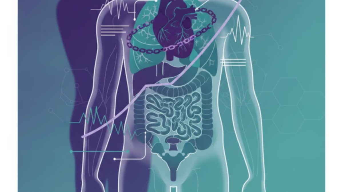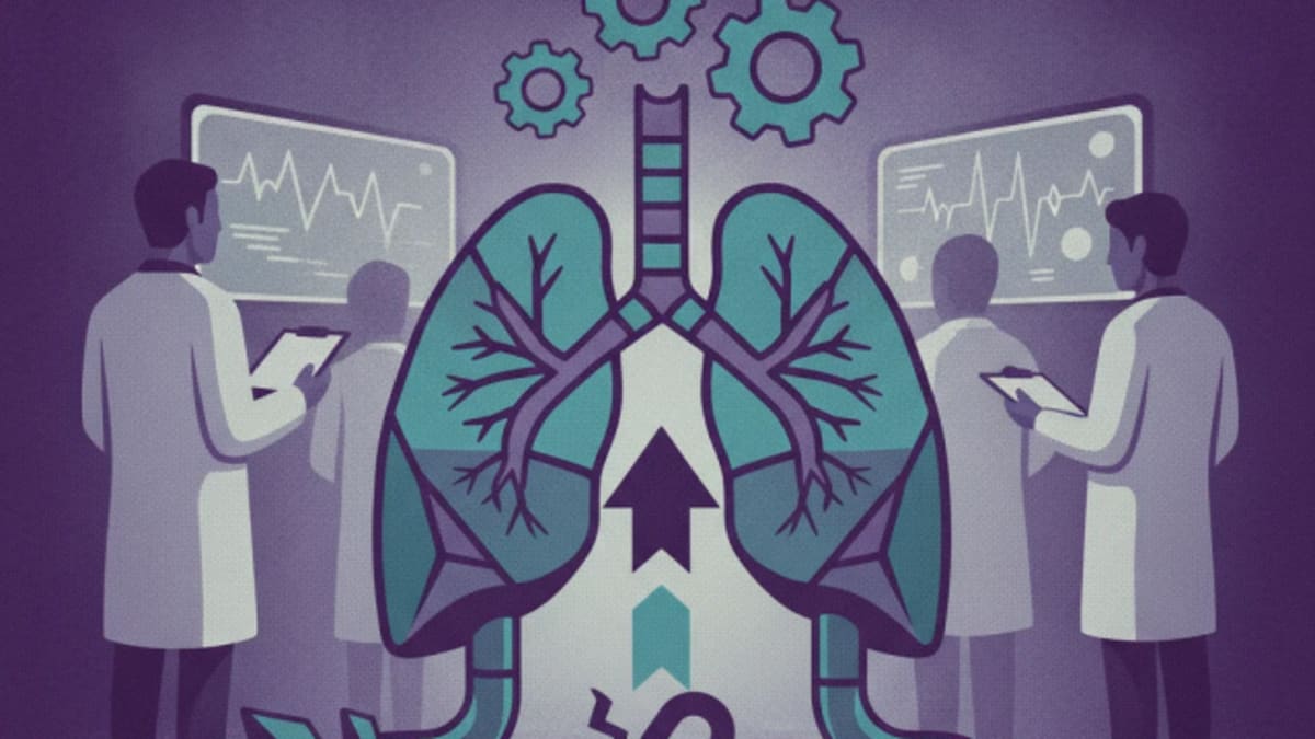

Duchenne muscular dystrophy (DMD) is a devastating X-linked genetic disorder, primarily affecting males, characterised by progressive muscle degeneration and weakness. While the skeletal muscle manifestations often dominate the early clinical picture, it is the insidious progression of cardiomyopathy and respiratory insufficiency that ultimately dictates prognosis and survival. These cardiac and pulmonary complications are not merely secondary effects; they are the leading causes of mortality in DMD patients, making vigilant surveillance a vital component of their long-term care.
Duchenne muscular dystrophy results from mutations in the DMD gene, leading to the absence or dysfunction of dystrophin, a protein vital for muscle cell integrity. Without functional dystrophin, muscle fibres are highly susceptible to damage during contraction, leading to chronic inflammation, fibrosis, and progressive muscle loss. This process affects not only skeletal muscles but also the myocardium and respiratory muscles, albeit with different pathological timelines and clinical presentations. The heart muscle, particularly the left ventricle, undergoes a similar degenerative process, leading to dilated cardiomyopathy, which often manifests silently until advanced stages. Similarly, the diaphragm and intercostal muscles weaken, impairing ventilation and cough effectiveness, predisposing patients to respiratory infections and failure.
The natural history of DMD has seen a dramatic shift over the past few decades, largely due to advancements in corticosteroid therapy, orthopaedic management, and, importantly, the systematic implementation of cardiac and respiratory surveillance and intervention. Historically, patients rarely survived beyond their late teens or early twenties, with cardiorespiratory failure being the inevitable endpoint. Today, with comprehensive care, many individuals with DMD live into their thirties and beyond, demonstrating the profound impact of proactive management. This extended survival, however, brings with it a greater need for sophisticated and sustained monitoring of the organ systems most vulnerable to dystrophin deficiency.
The Cardiac Imperative: Early Detection and Intervention
Cardiac involvement in DMD, primarily dilated cardiomyopathy, is almost universal, affecting nearly all patients by adulthood. The challenge lies in its often asymptomatic nature in early stages. Patients may maintain normal ejection fractions for years, even as subclinical myocardial damage progresses. This silent progression necessitates a proactive surveillance strategy, beginning from the time of diagnosis, typically in early childhood. The current standard of care recommends regular cardiac evaluations, even in the absence of symptoms, to detect early signs of myocardial dysfunction and initiate cardioprotective therapies before irreversible damage occurs.
Echocardiography remains the cornerstone of cardiac surveillance. Guidelines recommend baseline echocardiograms at diagnosis or by age 6, followed by annual assessments. These evaluations focus on left ventricular ejection fraction (LVEF), chamber dimensions, and wall motion abnormalities. Early changes, such as subtle reductions in global longitudinal strain, may precede overt LVEF decline, offering a window for earlier intervention. Cardiac magnetic resonance imaging (CMR) provides a more detailed assessment of myocardial fibrosis and inflammation, often detecting pathology before echocardiography. CMR is particularly valuable for characterising myocardial tissue and quantifying ventricular function with high precision, and it is increasingly incorporated into surveillance protocols, especially when echocardiographic findings are equivocal or when considering advanced therapies. For a deeper understanding of cardiac conditions, the Braunwald's Heart Disease, 12th Edition textbook offers comprehensive insights.
The initiation of cardioprotective medications, specifically angiotensin-converting enzyme (ACE) inhibitors and beta-blockers, is a critical intervention. These agents are typically started when there is evidence of left ventricular dysfunction, even if asymptomatic, or sometimes even prophylactically in older patients with normal function but significant fibrosis on CMR. ACE inhibitors, such as perindopril or enalapril, are often the first-line choice, aiming to reduce cardiac remodelling and improve ventricular function. Beta-blockers, like carvedilol, are added, particularly if there is persistent dysfunction or signs of heart failure. The timing of these interventions is paramount; delaying treatment until symptomatic heart failure develops significantly limits their efficacy and worsens prognosis. Regular electrocardiograms (ECGs) are also part of the surveillance, typically performed annually, to monitor for arrhythmias and conduction abnormalities, which can become more prevalent as cardiomyopathy progresses.
Navigating Respiratory Decline: From Ventilation to Cough Assistance
Respiratory muscle weakness in DMD progresses in parallel with skeletal muscle decline, leading to restrictive lung disease, hypoventilation, and impaired cough. This makes patients highly susceptible to respiratory infections, atelectasis, and ultimately, respiratory failure. Like cardiac involvement, respiratory compromise often progresses silently, with patients adapting to chronic hypoventilation until symptoms become severe. Therefore, systematic respiratory surveillance is as vital as cardiac monitoring, aiming to anticipate and mitigate these complications.
Pulmonary function tests (PFTs) are the primary tools for monitoring respiratory status. Forced vital capacity (FVC) is a key measure, typically assessed every 6 to 12 months, starting from around age 6. A decline in FVC, particularly when it falls below 30% of predicted, signals significant respiratory muscle weakness and often correlates with the need for ventilatory support. Peak cough flow (PCF) is another critical parameter, as an ineffective cough significantly increases the risk of aspiration and respiratory infections. A PCF below 160 L/min indicates impaired cough, necessitating mechanical cough assistance. Nocturnal hypoventilation often precedes daytime symptoms, making overnight oximetry or polysomnography important tools for early detection. These studies can identify periods of desaturation and hypercapnia during sleep, prompting the initiation of nocturnal ventilatory support.
Non-invasive ventilation (NIV), typically delivered via bilevel positive airway pressure (BiPAP), is a life-extending intervention for DMD patients with respiratory insufficiency. It is usually initiated when there is evidence of nocturnal hypoventilation, symptomatic daytime hypoventilation, or a significant decline in FVC. The goal of NIV is to improve alveolar ventilation, reduce the work of breathing, and prevent respiratory muscle fatigue. Patients require careful titration of ventilatory settings and ongoing monitoring to ensure optimal support. Mechanical insufflation-exsufflation devices, commonly known as cough assist devices, are essential for patients with impaired cough. These devices help clear airway secretions, reducing the risk of pneumonia and atelectasis, and are particularly important during respiratory infections. Clinicians should also consider the broader context of respiratory health, including vaccination status for influenza and pneumococcus, and prompt treatment of respiratory infections. For more on managing respiratory challenges, the Oxford Handbook of Respiratory Medicine provides a concise reference.
The Multidisciplinary Approach and Unmet Needs
Effective management of DMD requires a truly multidisciplinary team, integrating paediatricians, neurologists, cardiologists, pulmonologists, physical therapists, occupational therapists, dietitians, and social workers. This coordinated approach ensures that all aspects of the disease, from motor function to psychosocial support, are addressed. Regular team meetings and clear communication channels are vital to harmonise care plans and adapt them as the disease progresses. The transition from paediatric to adult care is a particularly vulnerable period, requiring careful planning to ensure continuity of surveillance and management. This is a common challenge across many chronic paediatric conditions, as highlighted in our coverage of myelofibrosis transplant timing, where early referral and coordinated care are similarly critical for long-term outcomes.
Despite significant progress, unmet needs remain. The optimal timing for initiating cardioprotective medications, particularly prophylactic use in patients with normal LVEF but early fibrosis, continues to be an area of active discussion. The long-term impact of newer genetic therapies on cardiac and respiratory outcomes is also still being elucidated. Access to specialised multidisciplinary care and advanced diagnostic tools, such as CMR, varies significantly across regions, creating disparities in patient outcomes. The psychological burden on patients and their families, managing a progressive and life-limiting illness, also requires ongoing attention and support, often overlooked in the focus on physical parameters.
The open-label nature of many observational studies informing current guidelines is an obvious caveat. While randomised controlled trials are challenging in rare diseases like DMD, the evidence base for surveillance benefits largely stems from cohort studies and expert consensus. The heterogeneity in disease progression among patients, even with similar genetic mutations, means that surveillance schedules must be individualised, while still adhering to general principles. The reliance on patient cooperation for PFTs and adherence to NIV also presents practical challenges, particularly in younger children or those with significant cognitive impairment. These factors underscore the need for flexible, patient-centred care models that can adapt to individual needs and circumstances.
Still, the evidence is clear: consistent, guideline-driven cardiac and respiratory surveillance, coupled with timely interventions, has transformed the prognosis for individuals with Duchenne muscular dystrophy. It has shifted the narrative from inevitable early mortality to one of extended survival and improved quality of life, allowing patients to live longer, more fulfilling lives. The ongoing challenge is to ensure that these established best practices are universally accessible and consistently applied, bridging the gap between knowledge and implementation in every clinic and community.
The message for clinicians managing Duchenne muscular dystrophy is unequivocal: cardiac and respiratory surveillance is not optional, it is foundational. Waiting for symptoms to emerge before initiating monitoring or intervention is a dereliction of modern care standards. These patients will develop cardiomyopathy and respiratory failure; the only variable is when and how aggressively it is managed.
GPs and specialists alike must integrate these surveillance schedules into their routine practice from the point of diagnosis. This means annual echocardiograms and pulmonary function tests, with low thresholds for referral to cardiology and pulmonology. The proactive initiation of ACE inhibitors and beta-blockers, even in asymptomatic patients with early signs of cardiac dysfunction, can genuinely alter the disease trajectory. Similarly, early consideration of non-invasive ventilation and cough assist devices, guided by objective measures like FVC and PCF, prevents acute crises and prolongs life.
The industry must continue to invest in developing therapies that target the underlying dystrophin deficiency, but also in improving diagnostic tools and access to care. While genetic therapies hold promise, the immediate impact on survival still rests heavily on meticulous supportive care. Ensuring that every patient has access to a coordinated multidisciplinary team, regardless of their geographical location, should be a priority for healthcare systems.
The survival gains in DMD are a demonstration of what systematic, evidence-based supportive care can achieve. It is a reminder that sometimes, the most impactful interventions are not novel drugs, but the consistent application of established guidelines. The schedule that changes survival is the one that is followed rigorously.
- The Pivot Proactive, guideline-driven cardiac and respiratory surveillance shifts DMD management from reactive symptom control to preventative intervention, directly extending life expectancy.
- The Data Early and consistent use of cardioprotective agents and respiratory support, guided by surveillance, can significantly delay disease progression and improve quality of life.
- The Action Clinicians must implement comprehensive, multidisciplinary surveillance protocols from diagnosis, integrating cardiology and pulmonology into routine DMD care.
ART-2026-1676
·09/26
Drafted with AI assistance, reviewed and approved by the editorial team. This publication is intended for healthcare professionals, researchers, and life science industry professionals. Content is provided for informational and educational purposes only and does not constitute medical advice.

Digital health and patient experience are my beat: the apps, the wearables, the real-world evidence claims, and whether any of it changes outcomes. Sceptical by training and optimistic by instinct.
Cite This Article
Ward S, Voss M. Duchenne muscular dystrophy: why silent cardiac decline costs lives. The Life Science Feed. Published September 14, 2026. Updated September 14, 2026. Accessed September 14, 2026. https://thelifesciencefeed.com/rare-diseases/duchenne-muscular-dystrophy/practice/duchenne-muscular-dystrophy-why-silent-cardiac-decline-costs-lives.
Editorial & AI Standards
All content is researched from peer-reviewed, open-access sources: published trial data, clinical guidelines, and regulatory filings. AI tools are used solely to structure and summarise that evidence; no AI-generated conclusions appear without editor verification against the primary source.
Every article is reviewed by a named editor before publication. Source citations are listed in the References section. This content does not represent the views of any pharmaceutical company, medical device manufacturer, or healthcare provider.
Licence & Rights
© 2026 The Life Science Feed. All rights reserved. Unless otherwise indicated, all content is the property of The Life Science Feed and may not be reproduced, distributed, or transmitted in any form or by any means without prior written permission.
Medical Disclaimer
The information provided on The Life Science Feed is for educational and informational purposes only. It is not intended as a substitute for professional medical advice, diagnosis, or treatment. Always seek the advice of your physician or other qualified healthcare provider regarding any medical condition or treatment decision. Never disregard professional medical advice or delay in seeking it because of something you have read on this website.
References
1. James KA, Gralla J, Ridall LA, et al. Left ventricular dysfunction in Duchenne muscular dystrophy. Cardiol Young. 2020;30(2):171-176. doi:10.1017/S1047951119002610
2. Riguzzi P, Grover E, Schiava M, et al. Respiratory function in Becker muscular dystrophy: a comprehensive longitudinal study. J Neurol Neurosurg Psychiatry. 2026;97(5):456-463. doi:10.1136/jnnp-2025-337953
3. Frishman N, Conway KC, Andrews J, et al. Perceived quality of life among caregivers of children with a childhood-onset dystrophinopathy: a double ABCX model of caregiver stressors and perceived resources. Health Qual Life Outcomes. 2017;15(1):33. doi:10.1186/s12955-017-0612-1