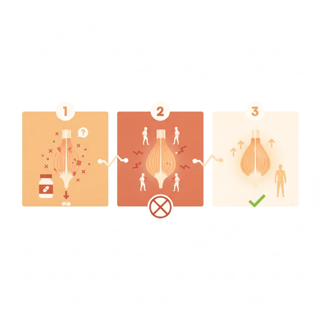

Statins remain the cornerstone of lipid-lowering therapy, demonstrably reducing cardiovascular morbidity and mortality across a broad spectrum of patients. Their widespread use, however, brings into focus the spectrum of potential adverse effects, from mild myalgia to severe myopathy. While most muscle-related symptoms are benign and resolve with statin discontinuation, a rare but clinically significant entity, statin-associated immune-mediated necrotising myopathy (IMNM), presents a distinct challenge for diagnosis and management.
This condition, unlike typical statin myopathy, involves an autoimmune process that necessitates specific immunomodulatory treatment beyond simple drug withdrawal. Understanding its unique pathophysiology and clinical presentation is important for clinicians to differentiate it from more common, less severe muscle complaints, as misdiagnosis can lead to prolonged muscle damage and functional impairment.
Statins are among the most commonly prescribed medications globally, lauded for their efficacy in primary and secondary prevention of atherosclerotic cardiovascular disease. Their mechanism of action involves inhibiting HMG-CoA reductase, the rate-limiting enzyme in cholesterol synthesis, leading to reduced hepatic cholesterol production and increased LDL receptor expression. This results in lower circulating LDL-C levels, a primary driver of cardiovascular risk. But while their benefits are undeniable, muscle-related adverse events, collectively termed statin-associated muscle symptoms (SAMS), are a frequent reason for discontinuation or dose reduction, affecting a significant proportion of patients.
SAMS encompass a range of symptoms, from mild myalgia and cramps to more severe myopathy, characterised by muscle weakness and elevated creatine kinase (CK) levels, and in rare cases, rhabdomyolysis. Most SAMS are dose-dependent, reversible upon statin withdrawal, and often attributed to direct mitochondrial toxicity or impaired coenzyme Q10 synthesis. The vast majority of patients experiencing SAMS will see their symptoms resolve within weeks of stopping the statin, or with a switch to a different statin or lower dose. But a small subset of patients develops a more insidious and persistent form of myopathy, which continues to progress even after statin cessation. This is the realm of statin-associated immune-mediated necrotising myopathy.
Distinguishing IMNM from typical SAMS
Immune-mediated necrotising myopathy, whether statin-associated or idiopathic, represents a severe autoimmune disorder characterised by extensive muscle fibre necrosis with minimal inflammatory cell infiltration on biopsy. The key differentiator for statin-associated IMNM is its temporal relationship with statin exposure and the presence of specific autoantibodies. Unlike typical SAMS, which usually improve rapidly after statin discontinuation, IMNM symptoms often persist or even worsen, demanding a different diagnostic and therapeutic approach. This distinction is paramount for clinicians, as misdiagnosis can lead to prolonged muscle damage and functional impairment.
The underlying mechanism of statin-associated IMNM involves an autoimmune response directed against 3-hydroxy-3-methylglutaryl-coenzyme A reductase (HMGCR), the very enzyme statins target. Statins are thought to unmask or trigger this autoimmune response in genetically predisposed individuals. The presence of anti-HMGCR antibodies is a hallmark of this condition, found in a substantial majority of affected patients. These antibodies are highly specific and serve as an important diagnostic biomarker, differentiating IMNM from other forms of myopathy, including other idiopathic inflammatory myopathies like polymyositis or dermatomyositis. The clinical presentation typically involves subacute onset of proximal muscle weakness, often severe, affecting both upper and lower extremities. Patients may report difficulty climbing stairs, rising from a chair, or lifting objects. Muscle pain may or may not be prominent, but objective weakness is consistently present. Creatine kinase levels are characteristically very high, often exceeding 1000 U/L, and can be significantly elevated even in the absence of severe pain, distinguishing it from simple myalgia. The persistence of these elevated CK levels and progressive weakness despite statin withdrawal is a strong indicator to investigate for IMNM.
Diagnostic pathways and confirmatory tests
When a patient presents with persistent or worsening myopathy after statin discontinuation, the diagnostic workup for IMNM should be initiated. Initial steps include a thorough history, physical examination focusing on muscle strength, and laboratory tests. Measuring CK levels is fundamental; persistently elevated levels, particularly those in the thousands, should raise a red flag. Testing for anti-HMGCR antibodies is the next important step. These antibodies can be detected in serum using various assays, including ELISA or immunoprecipitation. A positive anti-HMGCR antibody test in the context of statin exposure and persistent myopathy is highly suggestive of IMNM.
Muscle biopsy remains the gold standard for definitive diagnosis. Biopsy findings in IMNM are distinct: they show extensive myofiber necrosis and regeneration, with sparse or absent inflammatory infiltrates. This contrasts sharply with polymyositis, which features endomysial inflammation, or dermatomyositis, characterised by perifascicular atrophy and perivascular inflammation. Immunohistochemical staining may reveal sarcolemmal deposition of the membrane attack complex (C5b-9), further supporting an immune-mediated necrotising process. Electromyography (EMG) can also provide supportive evidence, typically showing myopathic changes with spontaneous activity such as fibrillation potentials and positive sharp waves, indicating active muscle damage. Nerve conduction studies are usually normal, helping to exclude neuropathic causes of weakness. For a comprehensive overview of diagnostic approaches in rheumatology, the Oxford Handbook of Rheumatology offers a concise reference.
Treatment strategies and prognosis
The management of statin-associated IMNM differs fundamentally from that of typical SAMS. Simple statin discontinuation is insufficient, as the autoimmune process continues to drive muscle damage. The cornerstone of treatment is immunosuppression. High-dose corticosteroids, such as oral prednisone, are typically initiated as first-line therapy. The goal is to rapidly suppress the autoimmune response and halt muscle necrosis. Patients often require prolonged courses of corticosteroids, with gradual tapering once clinical improvement and CK normalisation are achieved. But corticosteroids alone are often not enough, or their long-term use is limited by side effects. This is where steroid-sparing immunosuppressive agents come into play.
Commonly used steroid-sparing agents include methotrexate, azathioprine, mycophenolate mofetil, and rituximab. Intravenous immunoglobulin (IVIg) or plasma exchange (PLEX) may be used in severe or refractory cases, particularly in the acute phase to rapidly reduce antibody levels and inflammation. The choice of agent and duration of therapy are individualised, guided by disease severity, response to treatment, and patient tolerance. The treatment course is often protracted, lasting months to years, and requires close monitoring of muscle strength, CK levels, and antibody titres. Physical therapy and rehabilitation are also important components of management to help patients regain muscle strength and function. While many patients respond well to immunosuppressive therapy, some may experience residual weakness or require ongoing maintenance treatment. Early diagnosis and aggressive treatment are associated with better outcomes, highlighting the importance of recognising this rare but serious condition. This is a complex area, and the need for explicit protocols in managing such rare conditions is clear, as discussed in the real reason joint oncology practices need explicit protocols, a principle that extends to rare myopathies.
Unmet needs and future directions
Despite advances in understanding and managing IMNM, several challenges remain. The rarity of the condition means that awareness among general practitioners and even some specialists can be low, leading to diagnostic delays. There is no universally accepted treatment algorithm, and management often relies on expert opinion and extrapolation from other autoimmune myopathies. Long-term outcomes, including the risk of relapse and the optimal duration of immunosuppression, are not fully elucidated. Research is ongoing to identify additional biomarkers, refine diagnostic criteria, and develop more targeted therapies. Understanding the genetic predisposition to statin-associated IMNM is another area of active investigation, which could eventually lead to predictive screening tools. For now, vigilance and a high index of suspicion remain the most effective tools for clinicians.
The open-label nature of many case series and observational studies on IMNM is an obvious caveat. The lack of randomised controlled trials, while understandable given the rarity of the condition, means that treatment recommendations are largely based on expert consensus and retrospective data. This gap matters, as it leaves clinicians without robust, evidence-based comparisons of different immunosuppressive regimens. The long-term impact of chronic immunosuppression in this patient population, including risks of infection and malignancy, requires careful consideration and ongoing monitoring. The precise mechanism by which statins trigger the autoimmune response in genetically susceptible individuals also remains an area of active research, with implications for both prevention and targeted therapy development. The field continues to grapple with these complexities, much like the ongoing debate around whether inflammation is the real enemy in persistent CAD, even after lipid optimisation.
The differential diagnosis for proximal muscle weakness and elevated CK is broad, encompassing genetic myopathies, metabolic myopathies, infectious myositis, and other drug-induced myopathies. A careful and systematic approach is essential to avoid misdiagnosis. For instance, distinguishing IMNM from polymyositis or dermatomyositis without a muscle biopsy and antibody testing can be challenging, as all can present with similar clinical features. The presence of anti-HMGCR antibodies, however, provides a powerful tool for differentiation. The clinical course of IMNM is often more severe and protracted than other inflammatory myopathies, further highlighting the need for accurate and timely diagnosis. The role of imaging, such as muscle MRI, can also be supportive, showing muscle oedema and fatty infiltration, but it is not specific enough for definitive diagnosis. A combination of clinical suspicion, laboratory testing, and muscle biopsy is required to confirm the diagnosis and guide appropriate therapy. The importance of early recognition cannot be overstated, as delayed treatment can lead to irreversible muscle damage and significant functional disability. This condition is a stark reminder that even widely used and generally safe medications can, in rare instances, trigger severe and complex adverse events, demanding a thorough understanding from the prescribing clinician.
The recognition of statin-associated immune-mediated necrotising myopathy as a distinct clinical entity fundamentally changes how clinicians should approach persistent muscle symptoms in patients on statins. It is no longer sufficient to simply stop the statin and wait; if symptoms persist or worsen, particularly with high CK levels, a deeper investigation is warranted. This demands a shift in diagnostic algorithms, moving beyond the common SAMS framework to consider autoimmune pathology.
For patients, this means a more proactive and aggressive diagnostic workup, potentially leading to earlier initiation of immunosuppressive therapy. Delayed diagnosis can result in significant, irreversible muscle damage and prolonged disability. Clinicians must be prepared to refer to rheumatology or neurology specialists for antibody testing and muscle biopsy, rather than simply attributing all muscle complaints to benign statin intolerance.
The pharmaceutical industry, while focused on efficacy and common adverse events, also bears a responsibility to continue educating prescribers on rare but severe adverse drug reactions. Clearer guidelines and educational materials on differentiating IMNM from typical SAMS could significantly improve patient outcomes. This rare condition highlights the need for ongoing pharmacovigilance and a thorough understanding of drug-induced autoimmunity, even for well-established therapies.
- The Pivot Statin-associated IMNM is a distinct autoimmune myopathy, not merely an exacerbation of typical statin-induced muscle symptoms.
- The Data Patients typically present with proximal muscle weakness and elevated creatine kinase levels, often persisting or worsening after statin discontinuation.
- The Action Suspect IMNM in patients with persistent or worsening myopathy after statin cessation; confirm with muscle biopsy and autoantibody testing, then initiate immunosuppressive therapy.
ART-2026-1211
·10/26
Drafted with AI assistance, reviewed and approved by the editorial team. This publication is intended for healthcare professionals, researchers, and life science industry professionals. Content is provided for informational and educational purposes only and does not constitute medical advice.

Infectious disease, epidemiology, and global health equity. I have covered outbreaks from Ebola to COVID-19 to mpox. The stories I am most drawn to are where the science is clear and the response is slow.
Cite This Article
Reeves T, Voss M. Statin-associated myopathy: rare, real, and reversible?. The Life Science Feed. Published August 27, 2026. Updated October 5, 2026. Accessed October 12, 2026. https://thelifesciencefeed.com/rheumatology/inflammatory-myopathies/insights/statin-immune-mediated-necrotising-myopathy.
Editorial & AI Standards
All content is researched from peer-reviewed, open-access sources: published trial data, clinical guidelines, and regulatory filings. AI tools are used solely to structure and summarise that evidence; no AI-generated conclusions appear without editor verification against the primary source.
Every article is reviewed by a named editor before publication. Source citations are listed in the References section. This content does not represent the views of any pharmaceutical company, medical device manufacturer, or healthcare provider.
Licence & Rights
© 2026 The Life Science Feed. All rights reserved. Unless otherwise indicated, all content is the property of The Life Science Feed and may not be reproduced, distributed, or transmitted in any form or by any means without prior written permission.
Medical Disclaimer
The information provided on The Life Science Feed is for educational and informational purposes only. It is not intended as a substitute for professional medical advice, diagnosis, or treatment. Always seek the advice of your physician or other qualified healthcare provider regarding any medical condition or treatment decision. Never disregard professional medical advice or delay in seeking it because of something you have read on this website.
References
1. Li C, Liu H, Yang L, Liu R, Yin G, Xie Q. Immune-mediated necrotizing myopathy: A comprehensive review of the pathogenesis, clinical features, and treatments. J Autoimmun. 2024;148:103286. doi:10.1016/j.jaut.2024.103286
2. Suh J, Amato AA. Management of immune-mediated necrotizing myopathy. Muscle Nerve. 2024;70(2):166-172. doi:10.1002/mus.28114
3. Day JA, Limaye V. Immune-mediated necrotising myopathy: A critical review of current concepts. Semin Arthritis Rheum. 2019;49(3):420-429. doi:10.1016/j.semarthrit.2019.04.002
4. Khoo T, Chinoy H. Anti-HMGCR immune-mediated necrotising myopathy: Addressing the remaining issues. Autoimmun Rev. 2023;22(12):103468. doi:10.1016/j.autrev.2023.103468
5. Allenbach Y, Benveniste O, Stenzel W, Boyer O. Immune-mediated necrotizing myopathy: clinical features and pathogenesis. Nat Rev Rheumatol. 2020;16(12):689-701. doi:10.1038/s41584-020-00515-9
6. Pinal-Fernandez I, Casal-Dominguez M, Mammen AL. Immune-Mediated Necrotizing Myopathy. Curr Rheumatol Rep. 2018;20(4):21. doi:10.1007/s11926-018-0732-6









