
In busy clinical settings, the reliance on automated urinalysis reports is increasing. However, a recent case highlights how this practice can lead to missed critical diagnostic clues, delaying appropriate management for patients with acute kidney injury.
This case underscores the enduring value of manual urine microscopy, especially when initial dipstick results suggest significant renal abnormalities, guiding more precise and timely interventions.
The Case: A Diagnostic Impasse
A 62-year-old man with a history of hypertension presents to the emergency department with fatigue and decreased urine output over the past week. His medications include amlodipine and hydrochlorothiazide. Initial workup reveals a serum creatinine of 3.2 mg/dL (baseline 1.0 mg/dL), BUN of 45 mg/dL, and normal electrolytes. Urinalysis shows 2+ protein, 3+ blood, and the automated microscopy report indicates "few cellular elements, no casts seen." The emergency physician, faced with a busy department, initiates intravenous fluids and consults nephrology for possible acute kidney injury. But what's the cause?
Is it pre-renal azotemia from volume depletion? Is it intrinsic renal disease? Or is there an obstruction? Without a clear etiology, the patient is admitted for further evaluation. An ultrasound is ordered to rule out obstruction, but it comes back negative. The nephrologist, reviewing the case, notes the discrepancy between the significant hematuria and proteinuria on the dipstick and the automated microscopy report of "no casts." This prompts a request for manual urine microscopy.
The Microscopic Clue: Red Cell Casts
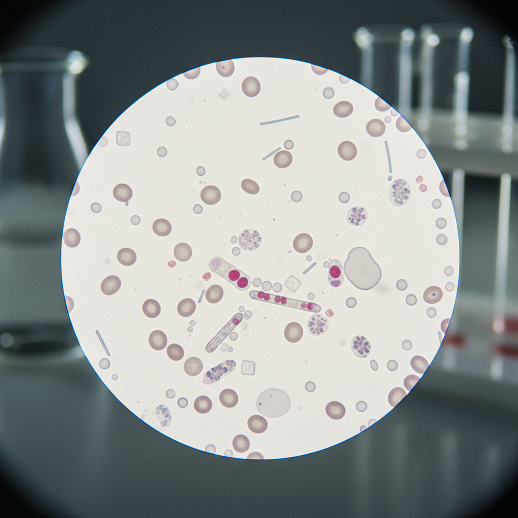
The manual microscopy reveals numerous red blood cell casts. These casts are pathognomonic for glomerular disease - specifically, glomerulonephritis. The automated analyzer, optimized for speed and cost-effectiveness, had missed these critical clues. The presence of red cell casts dramatically narrows the differential diagnosis, pointing towards an inflammatory or immune-mediated process within the glomeruli.
Based on this finding, further serological testing is performed, revealing elevated anti-glomerular basement membrane (anti-GBM) antibodies. The diagnosis is now clear: anti-GBM disease (Goodpasture's syndrome), a rapidly progressive glomerulonephritis that, if left untreated, can lead to end-stage renal disease and pulmonary hemorrhage.
Guideline Context: When to Look Beyond the Dipstick
The Kidney Disease Improving Global Outcomes (KDIGO) guidelines for glomerulonephritis emphasize the importance of early diagnosis and treatment to preserve kidney function. While the guidelines recommend a kidney biopsy for definitive diagnosis, they also highlight the role of non-invasive tests, including urinalysis, in guiding the initial evaluation. In this case, the presence of hematuria and proteinuria should have triggered a more thorough investigation, including manual microscopy, especially given the acute presentation. This finding directly influences the next steps: immunosuppressive therapy and potentially plasmapheresis to remove the pathogenic antibodies.
This case directly contradicts the reliance on solely automated urinalysis reports, a practice which is becoming more and more common in busy clinical settings. The KDIGO guidelines strongly recommend correlating clinical findings with laboratory results, a step that was initially overlooked in this case.
Limitations of the Evidence
The "evidence" here isn't a randomized controlled trial. It's a single case, albeit a compelling one. The broader "evidence" supporting the value of urine microscopy comes from decades of clinical experience and observational studies demonstrating its ability to detect important renal abnormalities. However, the sensitivity and specificity of urine microscopy can vary depending on the experience of the examiner and the quality of the sample. Furthermore, there is a lack of standardized protocols for urine microscopy, which can lead to inter-laboratory variability. Is this reproducible across different labs? Who is paying for the training? How do we standardize reporting?
It is also critical to acknowledge the limitations of automated urine analyzers. While they are highly efficient at detecting common abnormalities, they may miss subtle or atypical findings, such as dysmorphic red blood cells or specific types of casts. The algorithms used by these analyzers are constantly evolving, but they are not always perfect, and they can be particularly unreliable in complex cases.
The Changing Landscape of Lab Medicine
The increasing consolidation of laboratory services and the pressure to reduce costs have led to a decline in the availability of experienced microscopists. Many hospitals now rely on automated analyzers for routine urinalysis, with manual microscopy reserved for only the most complex cases. This trend raises concerns about the potential for missed diagnoses and delayed treatment, particularly in patients with atypical or subtle renal diseases.
In an era where urinalysis is increasingly automated, this case serves as a stark reminder of the enduring value of a classic test: urine microscopy. It requires expertise, time, and a willingness to look beyond the numbers. But the diagnostic yield can be substantial, potentially altering the course of a patient's life. Do we risk losing this skill in favor of efficiency? And at what cost?
The most striking consequence of over-reliance on automated urinalysis is delayed diagnosis and potentially irreversible organ damage. This case of anti-GBM disease highlights the critical gap. Automated systems, while efficient, can miss pathognomonic findings like red cell casts. This omission directly impacts patient outcomes, as timely intervention with immunosuppressive therapy and plasmapheresis is crucial for preserving kidney function in rapidly progressive glomerulonephritis. The initial misstep here underscores a systemic issue in modern diagnostics.
For clinicians, this means a renewed emphasis on correlating clinical presentation with laboratory data. The KDIGO guidelines already stress this, but the pressure of busy departments can lead to shortcuts. We must question automated reports, especially when significant dipstick findings like gross hematuria and proteinuria are downplayed. Industry, particularly manufacturers of automated analyzers, must acknowledge these limitations. While algorithms improve, they are not infallible. Investment in training and standardization for manual microscopy is also essential, ensuring reproducibility across different laboratories.
Patients bear the brunt of these diagnostic oversights. A missed diagnosis of glomerulonephritis can lead to end-stage renal disease, requiring dialysis or transplantation. This is a significant burden on individuals and healthcare systems. While the evidence here is a single compelling case, decades of clinical experience support the value of manual urine microscopy. We need to ensure that cost-effectiveness does not compromise diagnostic accuracy, particularly in conditions where early intervention is paramount.
- The Pivot Manual urine microscopy can reveal critical diagnostic clues, like red cell casts, that automated analyzers may miss, significantly narrowing the differential diagnosis for acute kidney injury.
- The Data Red blood cell casts are pathognomonic for glomerular disease, specifically glomerulonephritis.
- The Action In cases of unexplained hematuria and proteinuria, especially with acute kidney injury, clinicians should request manual urine microscopy to avoid missing key diagnostic indicators.
ART-2026-1794
·09/26
Drafted with AI assistance, reviewed and approved by the editorial team. This publication is intended for healthcare professionals, researchers, and life science industry professionals. Content is provided for informational and educational purposes only and does not constitute medical advice.

Digital health and patient experience are my beat: the apps, the wearables, the real-world evidence claims, and whether any of it changes outcomes. Sceptical by training and optimistic by instinct.
Cite This Article
Ward S, Voss M. The subtle clues urine microscopy can still offer. The Life Science Feed. Published September 28, 2026. Updated September 28, 2026. Accessed September 29, 2026. https://thelifesciencefeed.com/nephrology/acute-kidney-injury/case/the-subtle-clues-urine-microscopy-can-still-offer.
Editorial & AI Standards
All content is researched from peer-reviewed, open-access sources: published trial data, clinical guidelines, and regulatory filings. AI tools are used solely to structure and summarise that evidence; no AI-generated conclusions appear without editor verification against the primary source.
Every article is reviewed by a named editor before publication. Source citations are listed in the References section. This content does not represent the views of any pharmaceutical company, medical device manufacturer, or healthcare provider.
Licence & Rights
© 2026 The Life Science Feed. All rights reserved. Unless otherwise indicated, all content is the property of The Life Science Feed and may not be reproduced, distributed, or transmitted in any form or by any means without prior written permission.
Medical Disclaimer
The information provided on The Life Science Feed is for educational and informational purposes only. It is not intended as a substitute for professional medical advice, diagnosis, or treatment. Always seek the advice of your physician or other qualified healthcare provider regarding any medical condition or treatment decision. Never disregard professional medical advice or delay in seeking it because of something you have read on this website.