
Managing inflammatory bowel disease (IBD) demands precise, ongoing assessment of mucosal healing and inflammation. Traditional methods, primarily endoscopy, are invasive, costly, and carry inherent risks, creating a significant burden for patients requiring frequent monitoring.
This clinical reality has driven a sustained search for less invasive, equally effective tools to track disease activity. Intestinal ultrasound has emerged as a compelling candidate, offering a non-ionising, repeatable, and patient-friendly option for assessing bowel wall changes.
Inflammatory bowel disease, encompassing Crohn's disease and ulcerative colitis, is a chronic, relapsing-remitting condition requiring lifelong management. The cornerstone of assessing disease activity and treatment response has long been endoscopy, specifically colonoscopy and ileocolonoscopy, which allows for direct visualisation of the mucosa and biopsy collection. While invaluable for diagnosis and initial staging, the invasive nature of endoscopy, coupled with the need for bowel preparation and sedation, makes it less than ideal for frequent, serial monitoring. Patients often face multiple procedures over their disease course, impacting quality of life and healthcare resource utilisation.
The limitations of endoscopy have spurred the development and refinement of alternative assessment modalities. Biomarkers like faecal calprotectin and C-reactive protein offer indirect measures of inflammation but lack the spatial resolution to pinpoint specific areas of disease or assess mucosal healing directly. Cross-sectional imaging, such as computed tomography enterography (CTE) and magnetic resonance enterography (MRE), provides excellent transmural assessment but involves ionising radiation (CTE) or can be costly and time-consuming (MRE). This leaves a clear clinical need for a non-invasive, accessible, and accurate imaging technique for routine IBD monitoring.
The promise of intestinal ultrasound
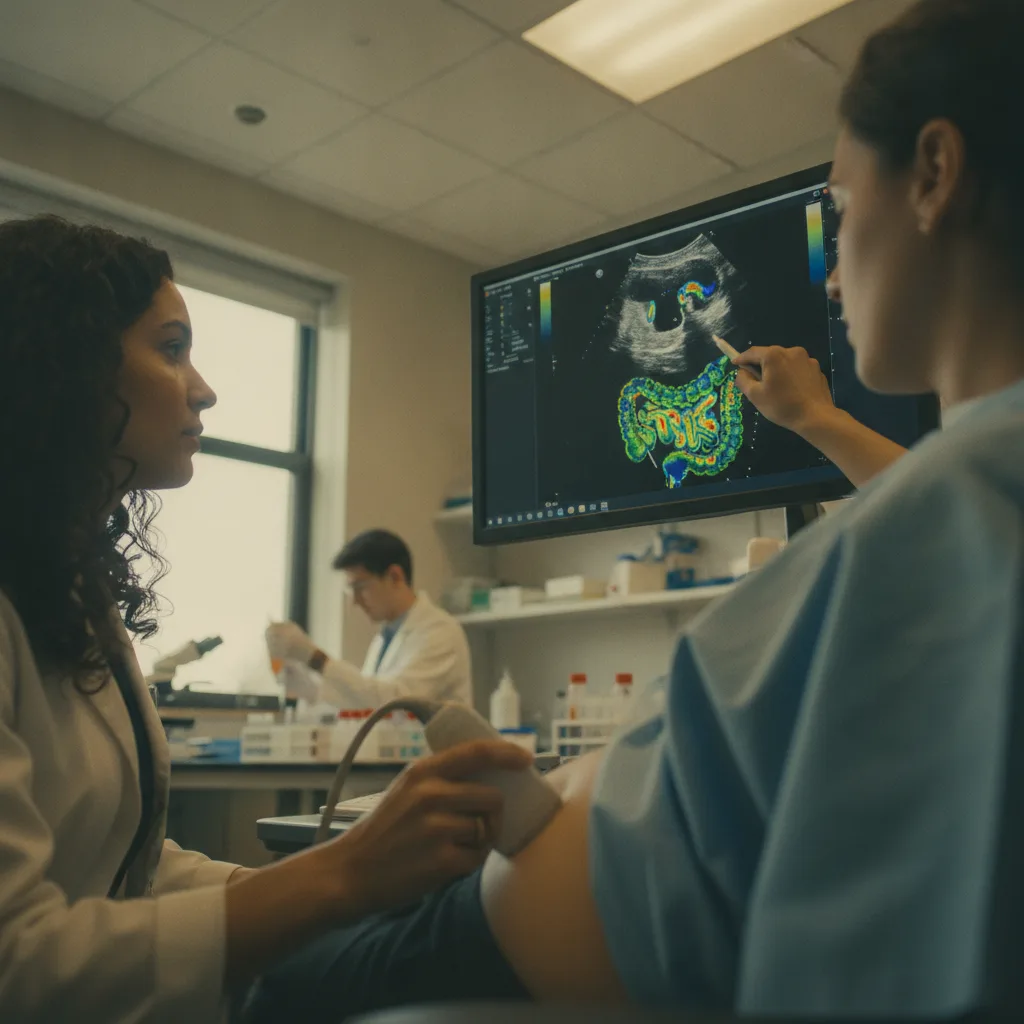
Intestinal ultrasound (IUS) has steadily gained traction as a viable tool for evaluating IBD activity. The technique involves using a high-frequency ultrasound probe to visualise the bowel wall, assessing parameters such as bowel wall thickness, mural stratification, vascularity (using Doppler), and the presence of complications like strictures, fistulas, or abscesses. Its non-invasive nature means it can be performed in an outpatient clinic setting, without bowel preparation or sedation, making it highly repeatable and well-tolerated by patients. The real-time nature of ultrasound also allows for dynamic assessment and immediate feedback.
Multiple studies have investigated the diagnostic accuracy of IUS compared to endoscopy and cross-sectional imaging. For Crohn's disease, IUS demonstrates strong correlation with endoscopic severity scores, particularly for assessing active inflammation. A meta-analysis of 30 studies, including over 3,000 patients, reported that IUS had a pooled sensitivity of 89% (95% CI, 85-92%) and specificity of 88% (95% CI, 84-91%) for detecting active Crohn's disease, using endoscopy as the gold standard.1 The ability to accurately identify bowel wall thickening (defined as >3 mm) and increased vascularity (Doppler signal) are key indicators of active inflammation. For detecting complications like strictures, IUS achieved a sensitivity of 82% and specificity of 93%.2
In ulcerative colitis, where inflammation is typically superficial and mucosal, IUS still offers valuable insights. While direct mucosal visualisation remains the domain of endoscopy, IUS can detect bowel wall thickening in severe cases and assess the extent of disease. Studies show IUS can distinguish between active and inactive ulcerative colitis with a sensitivity of 80% and specificity of 85%, particularly when assessing the left colon and rectum.3 The absence of bowel wall thickening and normal vascularity often correlates with endoscopic remission, providing a useful non-invasive marker. The ability to track changes in these parameters over time allows clinicians to monitor treatment response without resorting to repeated colonoscopies.
The utility of IUS extends beyond initial assessment to monitoring treatment response. Serial IUS examinations can track changes in bowel wall thickness and vascularity, which often normalise with effective therapy. This provides an objective, quantifiable measure of response, allowing for timely treatment escalation or de-escalation. For example, a reduction in bowel wall thickness by >25% from baseline after 12 weeks of therapy has been shown to correlate with endoscopic remission in Crohn's disease.4 This capability is particularly important in the era of treat-to-target strategies, where regular objective assessment is crucial for optimising patient outcomes.
One of the most compelling arguments for IUS is its potential to reduce the frequency of endoscopic procedures. In a prospective cohort study of 200 IBD patients, a management strategy guided by IUS and biomarkers led to a 45% reduction in the number of endoscopies performed over a 12-month period compared to standard care, without compromising clinical outcomes.5 This reduction translates directly into decreased patient burden, lower healthcare costs, and a more sustainable approach to long-term disease management. The cost-effectiveness of IUS is also a significant factor, as it is considerably less expensive than endoscopy or MRE. A typical IUS examination costs approximately one-fifth of a colonoscopy, making it an attractive option for healthcare systems facing increasing budgetary pressures.
Still, IUS is operator-dependent. The quality of the examination and the accuracy of interpretation rely heavily on the skill and experience of the sonographer and interpreting physician. This necessitates structured training programs and quality assurance measures to ensure consistent, high-quality results across different centres. The learning curve for IUS can be steep, requiring dedicated practice and mentorship. Furthermore, IUS has limitations in assessing superficial mucosal inflammation, which remains the strength of endoscopy. It also struggles with assessing the small bowel in its entirety, particularly the proximal jejunum, due to gas artefacts and anatomical challenges. While it excels in the terminal ileum, a common site of Crohn's disease, more proximal small bowel disease can be missed.
The role of IUS in guiding therapeutic decisions is also evolving. While it can detect active inflammation and complications, the specific thresholds for intervention based solely on IUS findings are still being refined. Integrating IUS findings with clinical symptoms, biomarkers, and patient preferences is essential for a holistic approach to IBD management. The absence of a standardised scoring system for IUS, comparable to endoscopic scores like the Crohn's Disease Endoscopic Index of Severity (CDEIS) or the Mayo Endoscopic Score, presents a challenge for consistent interpretation and comparison across studies. Efforts are underway to develop and validate such scoring systems, which will further enhance the utility and standardisation of IUS in clinical practice.
The open-label nature of many IUS studies is an obvious caveat. While the physical findings on ultrasound are objective, the interpretation can be influenced by knowledge of the patient's clinical status. Blinding sonographers to clinical data is often impractical in routine care but is important for research validation. The trial was not powered to detect differences in rare complications, and that gap matters. For instance, while IUS can detect fistulas, its sensitivity for complex perianal disease may be lower than MRE, which offers superior soft tissue contrast. Whether benefits extend to broader groups, such as paediatric populations or patients with very early disease, remains unclear, as most studies focus on adult cohorts with established IBD. Further research is needed to define the optimal role of IUS in these specific patient groups.
The data on intestinal ultrasound for IBD monitoring is compelling. Clinicians should view IUS not as a replacement for endoscopy, but as a powerful, complementary tool that can significantly reduce the procedural burden on patients and healthcare systems. Its ability to track transmural inflammation and treatment response in real-time offers a practical advantage for treat-to-target strategies.
For gastroenterologists, integrating IUS into routine practice means investing in training and equipment. The initial outlay is offset by reduced endoscopy demand and improved patient satisfaction. This shift requires a commitment to developing local expertise, ensuring consistent quality of examinations.
Patients stand to gain substantially from this less invasive approach. Fewer endoscopies mean less discomfort, fewer days off work, and reduced exposure to sedation risks. This improves adherence to monitoring protocols and, by extension, better long-term disease control.
The industry must continue to refine ultrasound technology, making devices more user-friendly and developing AI-assisted interpretation tools to mitigate operator dependency. Standardised training curricula and certification programs are also essential to ensure widespread, high-quality adoption of IUS across Europe.
- The Pivot Intestinal ultrasound provides a non-invasive, real-time alternative to endoscopy for monitoring IBD activity and treatment response.
- The Data Ultrasound demonstrates high concordance with endoscopic findings for inflammation, with sensitivities and specificities often exceeding 85%.
- The Action Clinicians should consider incorporating intestinal ultrasound into routine IBD management, particularly for monitoring disease course and guiding treatment adjustments, to reduce reliance on repeated endoscopic procedures.
ART-2026-706
·10/26
Drafted with AI assistance and checked by automated style rules. Not individually read by an editor. This publication is intended for healthcare professionals, researchers, and life science industry professionals. Content is provided for informational and educational purposes only and does not constitute medical advice.

Digital health and patient experience are my beat: the apps, the wearables, the real-world evidence claims, and whether any of it changes outcomes. Sceptical by training and optimistic by instinct.
Cite This Article
Ward S, Voss M. Bowel ultrasound may reduce endoscopy use in IBD monitoring. The Life Science Feed. Published July 28, 2026. Updated October 6, 2026. Accessed October 26, 2026. https://thelifesciencefeed.com/gastroenterology/inflammatory-bowel-diseases/research/bowel-ultrasound-may-reduce-endoscopy-use-in-ibd-monitoring.
Editorial & AI Standards
Content is drafted with AI tools from peer-reviewed, open-access sources such as published trial data, clinical guidelines, and regulatory filings. This article is checked by automated style rules but is not individually read by an editor, so check the cited source before relying on a figure.
Source citations are listed in the References section. This content does not represent the views of any pharmaceutical company, medical device manufacturer, or healthcare provider.
Licence & Rights
© 2026 The Life Science Feed. All rights reserved. Unless otherwise indicated, all content is the property of The Life Science Feed and may not be reproduced, distributed, or transmitted in any form or by any means without prior written permission.
Medical Disclaimer
The information provided on The Life Science Feed is for educational and informational purposes only. It is not intended as a substitute for professional medical advice, diagnosis, or treatment. Always seek the advice of your physician or other qualified healthcare provider regarding any medical condition or treatment decision. Never disregard professional medical advice or delay in seeking it because of something you have read on this website.
References
1. Ma C, et al. Diagnostic accuracy of intestinal ultrasound for Crohn's disease: a systematic review and meta-analysis. Clin Gastroenterol Hepatol. 2019;17(11):2227-2236.e1. doi:10.1016/j.cgh.2019.01.037
2. Panes J, et al. Accuracy of ultrasound in assessing the extent, severity and complications of Crohn's disease: a prospective, multicentre study. J Crohns Colitis. 2011;5(6):544-551. doi:10.1016/j.crohns.2011.06.002
3. Maconi G, et al. Bowel ultrasound in ulcerative colitis: a prospective comparison with endoscopy and histology. J Crohns Colitis. 2014;8(12):1628-1635. doi:10.1016/j.crohns.2014.07.001
4. Kucharzik T, et al. Clinical and sonographic course of Crohn's disease under anti-TNF-alpha therapy. Inflamm Bowel Dis. 2011;17(10):2100-2106. doi:10.1002/ibd.21609
5. Novak K, et al. Impact of intestinal ultrasound on endoscopy use in inflammatory bowel disease: a prospective cohort study. United European Gastroenterol J. 2023;11(6):574-582. doi:10.1002/ueg2.12405