
Tauopathies, including Alzheimer's disease, feature the hallmark of tau hyperphosphorylation, a pathological modification of the microtubule-associated protein tau. This modification has long been linked to neurodegeneration, but the precise mechanisms by which it disrupts neuronal function have remained elusive. A new study published in eLife now clarifies how hyperphosphorylated tau impairs microtubule binding and perturbs organelle trafficking, offering a deeper understanding of early disease pathology.1
Tau, a neuronal microtubule-associated protein (MAP), plays a critical role in organizing the axonal cytoskeleton and regulating intracellular transport. In healthy neurons, tau binds to microtubules cooperatively, forming cohesive envelopes that control access to the microtubule lattice and modulate the activity of motor proteins and other MAPs. This organized structure is fundamental for maintaining efficient axonal transport, a process vital for neuronal health and function. But in neurodegenerative conditions like Alzheimer's disease, tau undergoes hyperphosphorylation, a modification that has long been implicated in disease progression, yet its direct impact on tau's structural and functional roles remained incompletely understood.1
David Beaudet, a researcher at the University of Washington, and colleagues investigated how disease-related tau perturbations affect tau dynamics and its function as a selective barrier to intracellular transport. They used tau phospho-variants both in vitro and in live neurons to dissect the molecular consequences of hyperphosphorylation. Their work focused on understanding how these changes impact lysosome transport, a critical degradative pathway often disrupted in neurodegenerative diseases.1
The Disruption of Tau Envelopes
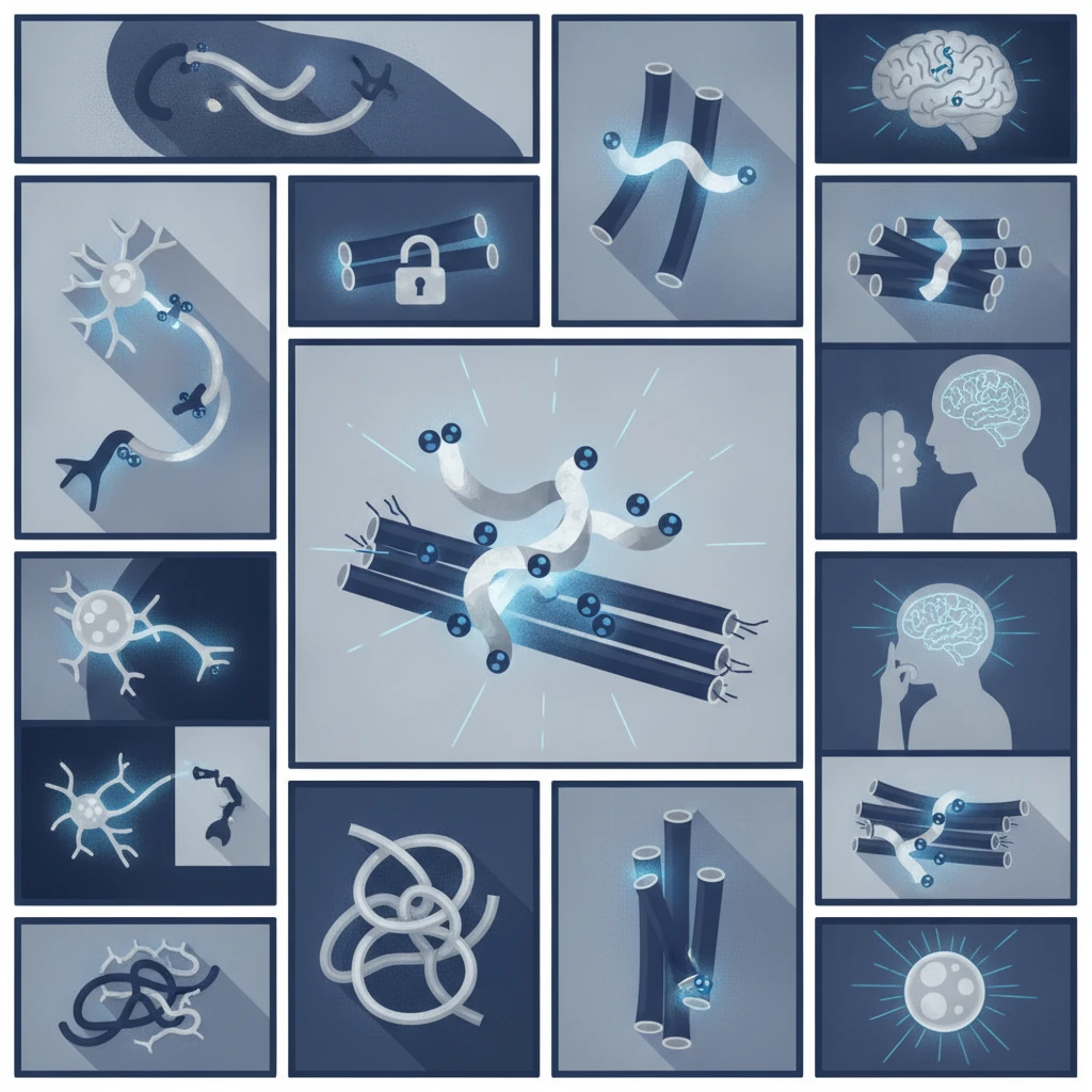
The study directly demonstrated that tau hyperphosphorylation fundamentally disrupts the cooperative binding of tau to microtubules. Wild-type tau forms distinct, cohesive envelopes around microtubules, creating a regulated environment for motor proteins and other MAPs. These envelopes are thought to act as a selective barrier, controlling the movement of cellular cargo along the microtubule tracks. The researchers observed this cooperative binding and envelope formation with unphosphorylated tau variants in their in vitro assays.1
But when tau was hyperphosphorylated, this cooperative binding mechanism failed. Hyperphosphorylated tau did not form these characteristic envelopes. Instead, it distributed more uniformly along the axon, suggesting a loss of its organized structure and function. This uniform distribution implies that the selective barrier function is compromised, potentially allowing unregulated access to the microtubule lattice. The researchers also found that hyperphosphorylated tau dissociated faster from microtubules, indicating a weaker and less stable interaction compared to its healthy counterpart. This reduced binding affinity and lack of envelope formation represent an early defect in tauopathies.1
This loss of structural integrity has profound implications for axonal transport. The cohesive tau envelope is not merely decorative; it is a functional component that modulates the interaction of motor proteins with microtubules. Without it, the finely tuned regulation of cargo movement is lost. The study's findings provide a clear mechanistic link between tau hyperphosphorylation and the structural disorganization of the axonal cytoskeleton, a hallmark of neurodegeneration.1
Understanding this initial disruption is important for developing therapies that aim to restore normal tau function. The precise molecular details of how hyperphosphorylation alters tau's interaction with microtubules offer specific targets for intervention. For instance, compounds that stabilize tau's cooperative binding or prevent hyperphosphorylation could potentially mitigate these early structural defects.1
Impact on Motor Protein Activity and Lysosome Transport
The study also meticulously examined how hyperphosphorylated tau affects the activity of two key motor proteins, KIF5C and KIF1A, which are responsible for transporting various organelles, including lysosomes, along microtubules. The researchers found that wild-type tau weakly inhibited KIF5C motility but strongly inhibited KIF1A. This differential inhibition suggests that healthy tau plays a role in regulating different motor proteins, possibly to fine-tune the transport of specific cargo.1
Hyperphosphorylation dramatically altered these inhibitory effects. For KIF5C, hyperphosphorylation reduced the inhibition exerted by tau. This means that KIF5C motors could move more freely along microtubules in the presence of hyperphosphorylated tau, potentially leading to increased or unregulated transport of its cargo. Conversely, for KIF1A, hyperphosphorylation increased inhibition. This heightened inhibition was attributed to a decrease in KIF1A processivity and an acceleration of its detachment from microtubules. In essence, hyperphosphorylated tau made it harder for KIF1A to move its cargo efficiently, causing it to stall or detach prematurely.1
These altered effects on motor proteins translated directly into observable changes in lysosome transport within live neurons. Lysosomes are essential for cellular waste removal and recycling, and their dysregulation is a common feature in neurodegenerative diseases. The researchers observed that phospho-resistant tau, which mimics healthy tau, inhibited processive lysosome motility, consistent with its role in forming a regulated barrier. But hyperphosphorylated tau weakened this tau-mediated regulation of lysosome transport.1
The effect of hyperphosphorylated tau on lysosome transport strikingly mimicked the observations in tau knockout neurons, which exhibit enhanced processivity of lysosomes. This suggests that hyperphosphoryylated tau not only loses its normal regulatory function but actively contributes to a state of dysregulation similar to having no tau at all. The enhanced processivity observed in tau knockout neurons, and now with hyperphosphorylated tau, indicates that lysosomes move more freely and perhaps less directionally, potentially leading to their mislocalization or inefficient delivery to degradation sites. This finding aligns with other research on AD biomarkers linked to worse cognition in midlife adults, where early cellular dysfunction precedes overt cognitive decline.1
The Clinical Relevance of Lysosome Dysregulation
The perturbation of lysosome trafficking by hyperphosphorylated tau is not merely a cellular curiosity; it has significant clinical implications. Lysosomes are critical for the degradation of misfolded proteins and damaged organelles, processes that are fundamental to neuronal health. When lysosome transport is impaired, these degradative pathways become inefficient, leading to the accumulation of toxic cellular waste. This accumulation is a well-established driver of neurodegeneration in tauopathies and other neurodegenerative diseases.1
Early defects in degradative pathways, such as those involving lysosomes, are thought to contribute to the initiation and progression of neurodegenerative processes. The study's findings provide a direct mechanistic link between tau hyperphosphorylation and these early defects. By disrupting the cooperative binding of tau to microtubules and subsequently dysregulating lysosome transport, hyperphosphorylated tau sets in motion a cascade of events that can lead to neuronal dysfunction and eventual cell death. This is particularly relevant given the broader context of neurodegenerative research, where understanding the initial triggers of pathology is paramount. The amyloid hypothesis, for example, has long dominated Alzheimer's research, but tau pathology is increasingly recognized as a critical, independent driver of neurodegeneration.1
The study used both in vitro models and live neurons, providing a robust platform for these observations. But the complexity of the neuronal environment in vivo, with its myriad interacting proteins and regulatory mechanisms, means that these findings represent a foundational step. Future research will need to validate these mechanisms in more complex animal models and ultimately in human tissue to fully understand their contribution to disease progression. The specific phosphorylation sites on tau that lead to these disruptive effects also warrant further investigation, as targeting these sites could offer therapeutic avenues. Clinicians looking for a comprehensive overview of neurological conditions might find the Oxford Handbook of Neurology (2nd ed) a useful quick-reference.1
The implications extend beyond Alzheimer's disease to other tauopathies, where similar mechanisms of tau pathology are at play. Understanding this common pathway of lysosome dysregulation could lead to broader therapeutic strategies applicable across a spectrum of neurodegenerative conditions. The study underscores the importance of targeting not just the aggregation of tau, but also the early functional consequences of its hyperphosphorylation, such as the disruption of microtubule binding and subsequent transport defects.1
What the Data Does Not Yet Establish
While the study provides compelling evidence for the direct impact of tau hyperphosphorylation on microtubule binding and lysosome transport, it does not fully elucidate the precise molecular machinery that mediates the cooperative binding of healthy tau. The mechanisms by which tau forms cohesive envelopes and how specific phosphorylation events disrupt these interactions at an atomic level remain areas for further exploration. The researchers used phospho-variants, but the full spectrum of disease-relevant phosphorylation patterns and their cumulative effects on tau function are complex and require more detailed structural and biochemical analyses.1
The study also primarily focused on lysosome transport. While critical, lysosomes are only one type of organelle transported along axons. Other vital cargoes, such as mitochondria, synaptic vesicles, and neurofilaments, are also transported by motor proteins like KIF5C and KIF1A. The differential effects of hyperphosphorylated tau on these other transport processes, and their combined contribution to neurodegeneration, were not within the scope of this particular investigation. A broader understanding of how hyperphosphorylated tau impacts the entire axonal transport system would provide a more complete picture of its pathological role.1
The study was conducted using in vitro systems and cultured neurons. While these models offer excellent control and allow for detailed mechanistic investigation, they do not fully replicate the complex environment of the living brain. Factors such as glial cell interactions, inflammatory responses, and the long-term effects of chronic tau pathology are not captured in these models. Translating these findings to human disease progression will require further studies in more physiologically relevant models, including animal models of tauopathy and post-mortem human brain tissue analysis. The long-term consequences of these transport defects on neuronal viability and network function also warrant further investigation.1
The study also did not explore potential compensatory mechanisms that neurons might employ in response to impaired lysosome trafficking. Cells are remarkably resilient, and it is possible that other pathways or proteins attempt to compensate for the dysregulation caused by hyperphosphorylated tau. Identifying and understanding these compensatory mechanisms could offer additional therapeutic targets. The findings are a significant step forward, but they open new avenues for research rather than closing the book on tau's complex pathology.1
The direct evidence that tau hyperphosphorylation dismantles cooperative microtubule binding and perturbs lysosome trafficking offers a clear, actionable target for intervention in tauopathies. For too long, the field has grappled with the downstream consequences of tau pathology, but this work points to a fundamental, early mechanism. Clinicians should recognize that the problem is not just tau aggregation, but the loss of its essential function as a regulator of intracellular transport.
This mechanistic clarity could guide the development of therapies that aim to restore tau's normal function, rather than simply clearing aggregated protein. Drugs designed to prevent specific phosphorylation events or to stabilize tau's interaction with microtubules could theoretically preserve lysosome function and, by extension, neuronal health. This shifts the focus from a purely symptomatic approach to one that addresses the root cause of cellular dysfunction.
But the challenge remains significant. Developing compounds that selectively modulate tau phosphorylation or binding without disrupting other critical cellular processes is complex. The differential effects on KIF5C and KIF1A highlight the intricate balance required. Any intervention must restore the delicate regulatory role of tau, not simply eliminate its pathological forms, which could lead to unintended consequences given tau's vital physiological functions.
These findings underscore the importance of early diagnosis and intervention in neurodegenerative diseases. If lysosome dysregulation is an early driver of neurodegeneration, then therapies targeting this pathway would likely be most effective when initiated before widespread neuronal damage occurs. This reinforces the ongoing push for better biomarkers and screening methods to identify individuals at the earliest stages of tauopathy.
- The Pivot Tau hyperphosphorylation directly disrupts its cooperative binding to microtubules, preventing the formation of cohesive envelopes essential for regulating intracellular transport.
- The Data Hyperphosphorylated tau did not form envelopes, distributed more uniformly along the axon, and dissociated faster from microtubules compared to wild-type tau.1
- The Action Understanding the precise dysregulation of lysosome transport by hyperphosphorylated tau offers a specific target for therapeutic intervention in tauopathies.
ART-2026-1902
·10/26
Drafted with AI assistance, reviewed and approved by the editorial team. This publication is intended for healthcare professionals, researchers, and life science industry professionals. Content is provided for informational and educational purposes only and does not constitute medical advice.

Infectious disease, epidemiology, and global health equity. I have covered outbreaks from Ebola to COVID-19 to mpox. The stories I am most drawn to are where the science is clear and the response is slow.
Cite This Article
Reeves T, Voss M. Tau: why its 'knockout' effect is a clue to alzheimer's. The Life Science Feed. Published October 13, 2026. Updated October 13, 2026. Accessed October 13, 2026. https://thelifesciencefeed.com/neurology/alzheimer-disease/research/tau-why-its-knockout-effect-is-a-clue-to-alzheimers.
Editorial & AI Standards
All content is researched from peer-reviewed, open-access sources: published trial data, clinical guidelines, and regulatory filings. AI tools are used solely to structure and summarise that evidence; no AI-generated conclusions appear without editor verification against the primary source.
Every article is reviewed by a named editor before publication. Source citations are listed in the References section. This content does not represent the views of any pharmaceutical company, medical device manufacturer, or healthcare provider.
Licence & Rights
© 2026 The Life Science Feed. All rights reserved. Unless otherwise indicated, all content is the property of The Life Science Feed and may not be reproduced, distributed, or transmitted in any form or by any means without prior written permission.
Medical Disclaimer
The information provided on The Life Science Feed is for educational and informational purposes only. It is not intended as a substitute for professional medical advice, diagnosis, or treatment. Always seek the advice of your physician or other qualified healthcare provider regarding any medical condition or treatment decision. Never disregard professional medical advice or delay in seeking it because of something you have read on this website.
References
1. Beaudet D, Berger CL, Hendricks AG. Tau hyperphosphorylation impairs cooperative binding to microtubules and perturbs organelle trafficking in neurons. Elife. 2026;15:e42804373. doi:10.7554/eLife.42804373