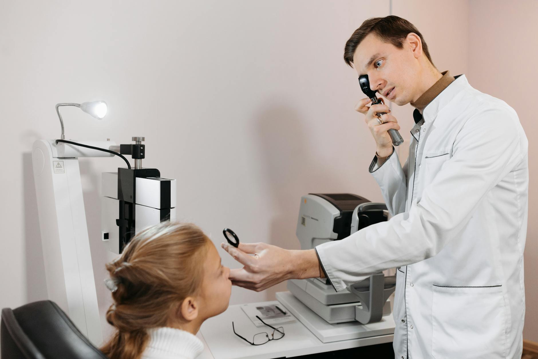
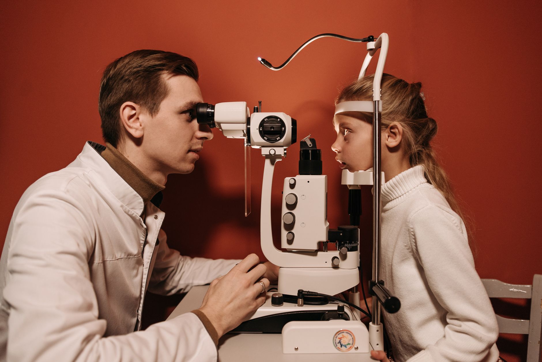
The eye has long been considered a window to the soul, but modern oculomics reveals it as a precise diagnostic portal to systemic health. Retinal imaging, once primarily for ophthalmic conditions, now offers a non-invasive glimpse into vascular, neurological, and metabolic states across the body. This evolving field promises to transform how general practitioners approach early disease detection and risk stratification.
The traditional ophthalmoscopic examination provides a limited view of the retina, often detecting systemic disease manifestations only at advanced stages. But the advent of high-resolution imaging technologies, such as optical coherence tomography (OCT), OCT angiography (OCTA), and ultrawide field imaging, has expanded this capability dramatically. These tools capture intricate details of the retinal vasculature, nerve fiber layers, and cellular structures, offering biomarkers for a range of non-ocular conditions. The retina, being an accessible part of the central nervous system, shares embryological origins and physiological characteristics with the brain, making it an ideal site for detecting systemic pathologies.
Oculomics, therefore, moves beyond simply diagnosing eye diseases. It aims to extract systemic health information from ocular images, using artificial intelligence (AI) and machine learning algorithms to process vast datasets. This approach allows for the identification of subtle patterns that are imperceptible to the human eye. The goal is to facilitate earlier diagnosis and intervention for conditions that might otherwise progress silently for years, impacting patient outcomes and healthcare costs.
The Retina as a Biomarker Hub
The retinal microvasculature provides a direct, non-invasive view of the body's small blood vessels, reflecting the systemic vascular health. Changes in retinal vessel caliber, tortuosity, and branching patterns are established indicators of hypertension, diabetes, and cardiovascular disease risk. For instance, narrowing of retinal arterioles and widening of venules correlate with increased blood pressure and a higher incidence of stroke and myocardial infarction. These microvascular alterations often precede the clinical onset of cardiovascular events, offering a critical window for intervention.
Diabetic retinopathy is a well-known ocular manifestation of diabetes, but oculomics extends this by identifying pre-clinical markers of metabolic dysfunction. Early changes in retinal blood flow, microaneurysms, and subtle exudates can be detected even before a formal diagnosis of diabetes. This allows for targeted lifestyle interventions or pharmacotherapy to prevent or delay the progression of both diabetes and its associated complications. The ability to monitor these changes over time provides a dynamic assessment of disease control and treatment efficacy, moving beyond static HbA1c measurements.
Beyond vascular changes, the retina's neural components offer insights into neurodegenerative diseases. The retinal nerve fiber layer (RNFL) and ganglion cell layer (GCL) are extensions of the central nervous system. Thinning of these layers, detectable by OCT, has been associated with conditions like Alzheimer's disease, Parkinson's disease, and multiple sclerosis. These changes can occur years before cognitive decline or motor symptoms become apparent, making the retina a potential site for early screening. The precision of OCT measurements allows for quantitative assessment of neural tissue loss, which can be tracked longitudinally to monitor disease progression or response to neuroprotective therapies. This is particularly relevant given the challenges in early diagnosis of neurodegenerative conditions, where current methods are often invasive or expensive.
Technical Advances Driving Oculomics
The rapid evolution of imaging technology underpins the expansion of oculomics. OCT, a non-invasive imaging technique, provides cross-sectional views of the retina with micron-level resolution. It allows for precise measurement of retinal layer thicknesses, which is essential for detecting subtle neural degeneration. OCTA, an extension of OCT, visualizes retinal microvasculature without the need for intravenous dye, offering detailed maps of blood flow and capillary density. This is invaluable for assessing vascular health and detecting early signs of conditions like diabetic retinopathy or retinal vein occlusion.
Ultrawide field imaging captures up to 200 degrees of the retina in a single shot, providing a comprehensive view of the peripheral retina, which is often affected by systemic diseases. This broader perspective allows for the detection of lesions or vascular changes that might be missed with traditional fundus photography. The integration of these imaging modalities with AI algorithms is where the true power of oculomics lies. AI models can analyze vast quantities of retinal images, identifying complex patterns and correlations that human clinicians might overlook. These algorithms can be trained to detect specific biomarkers for various systemic diseases, improving diagnostic accuracy and efficiency. For example, AI can quantify microaneurysms or identify subtle changes in vessel tortuosity with a consistency that is difficult for human graders to achieve.
The development of these AI-driven diagnostic tools is still in its early stages, but the potential for widespread clinical application is clear. These systems can process images rapidly, providing immediate feedback to clinicians. This could significantly reduce the burden on ophthalmologists and allow for more efficient screening in primary care settings. The ability to automate the detection of subtle changes means that even general practitioners could potentially use these tools to identify patients at risk, facilitating timely referrals to specialists. This shifts the paradigm from reactive disease management to proactive health monitoring, a concept also explored in cataract surgery considerations.
Integrating Oculomics into Clinical Practice
For general practitioners and specialists, the integration of oculomics into routine care presents both opportunities and challenges. The primary opportunity is the ability to screen for systemic diseases non-invasively and at an earlier stage. Imagine a scenario where a routine eye exam, perhaps even conducted in a community setting, could flag a patient for early signs of hypertension or a predisposition to Alzheimer's disease. This would enable earlier intervention, potentially delaying or preventing severe outcomes. The Oxford Handbook of Clinical Medicine provides a comprehensive overview of how such diagnostic insights can be integrated into broader patient management strategies.
But the challenges are substantial. The cost of advanced imaging equipment, the need for specialized training to interpret complex images, and the regulatory hurdles for AI-driven diagnostic tools are all significant barriers. Standardized protocols for image acquisition and interpretation are essential to ensure consistency and reliability across different clinical settings. The ethical implications of identifying disease risks years in advance, particularly for conditions with no current preventative therapies, must be carefully considered. How do clinicians communicate such information to patients, and what support systems need to be in place?
Still, the potential benefits outweigh these challenges. Oculomics could transform population health screening, allowing for targeted interventions in high-risk groups. It could also provide valuable insights for personalized medicine, tailoring treatment strategies based on an individual's unique risk profile as revealed by their retinal biomarkers. The field is moving towards point-of-care devices and telemedicine solutions, which could make advanced retinal imaging more accessible in remote or underserved areas. The ability to monitor disease progression and treatment response through non-invasive retinal scans could also reduce the need for more invasive or costly diagnostic procedures, improving patient comfort and reducing healthcare expenditures. This proactive approach contrasts with traditional methods, where conditions like dry eye disease are often diagnosed later, as discussed in recent findings on Xiidra.
Future Directions and Unanswered Questions
The future of oculomics involves refining AI algorithms to improve diagnostic accuracy and expand the range of detectable systemic diseases. Researchers are exploring the retina's potential as a biomarker for conditions beyond cardiovascular and neurodegenerative diseases, including kidney disease, autoimmune disorders, and even certain cancers. The integration of multi-modal data, combining retinal images with genetic information, blood biomarkers, and clinical history, will likely lead to even more powerful predictive models. This holistic approach could provide a comprehensive risk assessment for individual patients, guiding preventative strategies and personalized treatment plans.
But several questions remain. What is the optimal frequency for retinal screening in asymptomatic individuals? How do we ensure equitable access to these advanced technologies? And how will healthcare systems adapt to manage the influx of early diagnoses and the demand for preventative interventions? The validation of AI algorithms in diverse populations is also critical to ensure their generalizability and avoid biases. The clinical utility of these tools must be rigorously tested in large-scale prospective studies to demonstrate their impact on patient outcomes and healthcare costs. Without robust evidence, widespread adoption will be slow. The field needs to move beyond simply demonstrating correlations to proving that early detection via oculomics leads to tangible improvements in patient health. The next phase of research will focus on these implementation challenges, ensuring that oculomics fulfills its promise of ushering in a new era of proactive eye care.
The promise of oculomics is not merely incremental improvement; it is a fundamental shift in how we approach early disease detection. For general practitioners, this means the eye exam could evolve from a reactive diagnostic tool for vision problems to a proactive screening mechanism for systemic health. Identifying patients at risk for hypertension or neurodegeneration years in advance offers an unprecedented opportunity for preventative medicine, but it also demands new clinical pathways for managing these early signals.
The industry faces the challenge of developing affordable, user-friendly imaging devices and validated AI algorithms that can be seamlessly integrated into primary care. The current market of advanced retinal imaging often requires specialist equipment and interpretation, which limits its widespread utility. Simplifying these technologies and ensuring their accuracy and reliability across diverse patient populations will be essential for adoption.
For patients, oculomics offers the potential for earlier, less invasive diagnosis of serious conditions, allowing for interventions that could significantly improve their quality of life and longevity. But this also introduces the psychological burden of knowing about a future disease risk, especially for conditions with limited treatment options. Clinicians will need to develop new communication strategies to convey this information responsibly, ensuring patients understand both the benefits and the limitations of these early insights.
- The Pivot Oculomics leverages advanced retinal imaging to identify systemic disease markers before overt symptoms manifest.
- The Data Retinal microvascular changes correlate with cardiovascular risk, while specific retinal nerve fiber layer thinning associates with neurodegeneration.
- The Action Clinicians should consider the potential of integrating advanced retinal scans into routine health assessments for at-risk patients.
ART-2026-1864
·10/26
Drafted with AI assistance, reviewed and approved by the editorial team. This publication is intended for healthcare professionals, researchers, and life science industry professionals. Content is provided for informational and educational purposes only and does not constitute medical advice.

Thirty years in health journalism, the last fifteen in life sciences. I have reported from every major medical congress and watched blockbuster drugs get revised after approval. I cover what the data says.
Cite This Article
Carter J, Voss M. Oculomics: why your routine eye exam is missing critical clues. The Life Science Feed. Published October 2, 2026. Updated October 2, 2026. Accessed October 2, 2026. https://thelifesciencefeed.com/ophthalmology/cataract/innovation/oculomics-why-your-routine-eye-exam-is-missing-critical-clues.
Editorial & AI Standards
All content is researched from peer-reviewed, open-access sources: published trial data, clinical guidelines, and regulatory filings. AI tools are used solely to structure and summarise that evidence; no AI-generated conclusions appear without editor verification against the primary source.
Every article is reviewed by a named editor before publication. Source citations are listed in the References section. This content does not represent the views of any pharmaceutical company, medical device manufacturer, or healthcare provider.
Licence & Rights
© 2026 The Life Science Feed. All rights reserved. Unless otherwise indicated, all content is the property of The Life Science Feed and may not be reproduced, distributed, or transmitted in any form or by any means without prior written permission.
Medical Disclaimer
The information provided on The Life Science Feed is for educational and informational purposes only. It is not intended as a substitute for professional medical advice, diagnosis, or treatment. Always seek the advice of your physician or other qualified healthcare provider regarding any medical condition or treatment decision. Never disregard professional medical advice or delay in seeking it because of something you have read on this website.
References
1. Park JT, Tan YY, Ko YE, Thakur S, Rim TH, Joo YS. The Eye as a Window to Cardio-Kidney-Metabolic Disease. Clin J Am Soc Nephrol. 2026. doi:10.2215/CJN.0000001224
2. Wu JH, Liu TYA. Application of Deep Learning to Retinal-Image-Based Oculomics for Evaluation of Systemic Health: A Review. J Clin Med. 2022;12(1). doi:10.3390/jcm12010152
3. Ran AR, Zhu Z, Cheng KHL, et al. Artificial intelligence-based retinal imaging for brain health assessment: a scoping review. Lancet Digit Health. 2026;8(8):101020. doi:10.1016/j.landig.2026.101020
4. Bisen JB, Sikora H, Aneja A, Shah SJ, Mirza RG. Retinal Imaging as a Window into Cardiovascular Health: Towards Harnessing Retinal Analytics for Precision Cardiovascular Medicine. J Cardiovasc Dev Dis. 2025;12(6). doi:10.3390/jcdd12060230
5. Wallraven T, Günthner R, Ribeiro A, et al. Retinal vessel imaging in inflammatory disease: From endothelial dysfunction to clinical evidence and translation. Prog Retin Eye Res. 2026;112:101472. doi:10.1016/j.preteyeres.2026.101472
6. Wood K, Schnorbus N, Muncharaz L, et al. A novel electrocardiogram-synchronized laser Doppler holography system for cardiac cycle-resolved retinal hemodynamics: development and validation. Sci Rep. 2026;16(1). doi:10.1038/s41598-026-51137-2