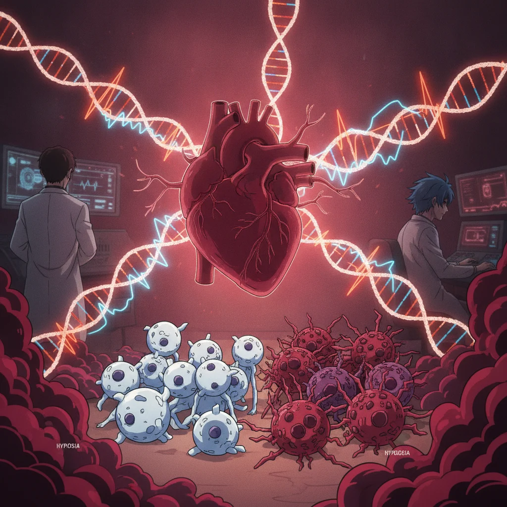

Endothelial cells are the gatekeepers of vascular health, forming a critical barrier between blood and tissue. When oxygen levels drop, a state known as hypoxia, these cells undergo profound changes that can either protect the vessel or contribute to disease. Understanding how hypoxia remodels gene expression in these cells is fundamental to grasping the pathogenesis of conditions from atherosclerosis to pulmonary hypertension.
The cistrome, the complete set of cis-acting regulatory elements in a genome, dictates which genes are turned on or off. In endothelial cells, the cistrome response to hypoxia is a complex relationship of transcription factors and epigenetic modifications, orchestrating a cellular adaptation that can have far-reaching consequences for cardiovascular health.
The human umbilical vein endothelial cell (HUVEC) is a widely used model for studying endothelial biology, offering a readily accessible and well-characterized system to investigate cellular responses to various stimuli, including hypoxia. These cells, derived from the umbilical cord vein, exhibit many characteristics of adult endothelial cells, making them relevant for understanding fundamental vascular processes. Hypoxia, a reduction in oxygen supply, is a common feature in numerous pathological states, including myocardial infarction, stroke, peripheral artery disease, and tumor microenvironments. The endothelial response to hypoxia is not merely a passive reaction; it is an active, coordinated genetic program designed to adapt the cell to low oxygen conditions, but this adaptation can also contribute to disease progression.
The cellular machinery responsible for sensing and responding to hypoxia is primarily centered around the hypoxia-inducible factor (HIF) family of transcription factors. HIFs are heterodimeric proteins composed of an oxygen-sensitive alpha subunit (HIF-1α, HIF-2α, or HIF-3α) and a constitutively expressed beta subunit (HIF-1β, also known as ARNT). Under normoxic conditions, HIF-α subunits are hydroxylated by prolyl hydroxylase domain (PHD) enzymes, leading to their ubiquitination by the von Hippel-Lindau (VHL) tumor suppressor protein and subsequent proteasomal degradation. But when oxygen is scarce, PHDs are inhibited, HIF-α subunits stabilize, translocate to the nucleus, and dimerize with HIF-1β. This complex then binds to hypoxia-response elements (HREs) in the promoters and enhancers of target genes, activating their transcription. The HIF pathway is a well-established master regulator of the hypoxic response, controlling genes involved in angiogenesis, erythropoiesis, glucose metabolism, and cell survival.
The cistrome's dynamic response to oxygen deprivation
The cistrome, the full complement of DNA regions bound by transcription factors and other chromatin-associated proteins, undergoes significant remodeling in response to hypoxia in HUVECs. This remodeling is not solely driven by HIFs. Other transcription factors, such as NF-κB, AP-1, and STAT3, also play roles, often interacting with HIFs or acting independently to regulate gene expression. The accessibility of chromatin, the tightly packed structure of DNA and proteins, is a critical determinant of gene expression. Hypoxia can induce changes in chromatin accessibility, making certain regulatory regions more or less available for transcription factor binding. This dynamic alteration of the epigenome is a key component of the cistrome response.
Chromatin immunoprecipitation sequencing (ChIP-seq) and Assay for Transposase-Accessible Chromatin using sequencing (ATAC-seq) are powerful techniques used to map transcription factor binding sites and open chromatin regions, respectively. Studies employing these methods have revealed that hypoxia leads to both opening and closing of chromatin at specific loci across the HUVEC genome. Regions associated with genes involved in angiogenesis, glycolysis, and inflammation often become more accessible, facilitating the binding of HIFs and other transcription factors. Conversely, some regions may become less accessible, leading to the downregulation of genes that are not essential or are detrimental under low oxygen conditions. This intricate dance of chromatin dynamics ensures a precise and tailored cellular response.
Transcription factor interplay and epigenetic modifications
While HIFs are central, their activity is not isolated. They interact with a multitude of other transcription factors, co-activators, and co-repressors to fine-tune gene expression. For instance, the interaction between HIF-1 and NF-κB is particularly relevant in inflammatory responses under hypoxia. NF-κB, a key regulator of immune and inflammatory genes, can be activated by hypoxic stress, and its activity can be modulated by HIF-1. This cross-talk, or relationship, can lead to a synergistic activation of genes involved in both angiogenesis and inflammation, contributing to the complex pathology seen in chronic hypoxic conditions.
Epigenetic modifications, such as DNA methylation and histone modifications, also play a significant role in shaping the cistrome response to hypoxia by influencing gene expression. DNA methylation, the addition of a methyl group to cytosine bases, typically leads to gene silencing when it occurs in promoter regions. Hypoxia can alter the activity of DNA methyltransferases and demethylases, leading to changes in methylation patterns. Similarly, histone modifications, including acetylation, methylation, and phosphorylation, can influence chromatin structure and gene accessibility. For example, histone acetylation generally promotes an open chromatin state, making genes more accessible for transcription. Hypoxia has been shown to affect the activity of histone acetyltransferases and deacetylases, leading to global and locus-specific changes in histone acetylation. These epigenetic changes provide an additional layer of regulatory complexity, allowing for sustained and heritable alterations in gene expression in response to prolonged hypoxic stress.
Implications for vascular disease and therapeutic targets
The cistrome response to hypoxia in HUVECs has significant implications for understanding and potentially treating various vascular diseases. In conditions like atherosclerosis, regions of plaque often experience chronic hypoxia. The endothelial cells lining these plaques respond by activating HIFs and other transcription factors, leading to increased expression of pro-inflammatory cytokines, adhesion molecules, and pro-angiogenic factors. These changes contribute to plaque growth, instability, and rupture. Understanding the specific cistrome alterations in these contexts could identify novel targets for therapeutic intervention. For example, inhibiting specific epigenetic enzymes that are aberrantly activated under hypoxia might normalize gene expression and mitigate disease progression.
Pulmonary hypertension is another condition where hypoxia plays a central role. Chronic hypoxia in the lungs leads to pulmonary vasoconstriction and vascular remodeling, driven in part by endothelial cell dysfunction. The cistrome changes in pulmonary artery endothelial cells under hypoxia are similar to those observed in HUVECs, involving HIF activation and epigenetic reprogramming. Targeting these pathways could offer new strategies for managing this debilitating disease. The Oxford Handbook of Cardiology provides a concise guide to modern cardiological practice, including discussions on pulmonary hypertension and its underlying mechanisms.
The open-label nature of many mechanistic studies is an obvious caveat; these are fundamental biological investigations, not clinical trials. The findings are derived from in vitro models, specifically HUVECs, which, while valuable, do not fully recapitulate the complexity of the human vascular system in vivo. The cellular environment, systemic factors, and interactions with other cell types in a living organism can significantly influence the hypoxic response. Whether the specific cistrome changes observed in HUVECs translate directly to endothelial cells in different vascular beds or in various disease states in humans remains an area for further investigation. The duration and severity of hypoxia also vary widely in pathological conditions, and the HUVEC model typically uses acute or sustained moderate hypoxia, which may not capture the full spectrum of physiological and pathological hypoxic challenges.
Still, the detailed mapping of transcription factor binding sites and chromatin accessibility provides a robust foundation for future research. The identification of specific HREs and other regulatory elements that are dynamically altered by hypoxia offers concrete targets for drug development. For instance, small molecules that selectively modulate the binding of HIFs or other key transcription factors to these regulatory regions could potentially normalize endothelial function under hypoxic stress. Understanding the epigenetic market changes, such as specific histone modifications or DNA methylation patterns, opens the door to epigenetic therapies that aim to reverse or prevent maladaptive gene expression. The field is moving towards a more comprehensive understanding of how these molecular events contribute to disease, paving the way for precision medicine approaches in vascular biology.
The detailed understanding of how hypoxia reshapes the endothelial cistrome in HUVECs provides a critical mechanistic foundation for vascular disease. Clinicians often manage the downstream consequences of chronic hypoxia, but this work points to upstream regulatory events that could be targeted. It suggests that interventions aimed at normalizing chromatin accessibility or specific transcription factor activity might offer a more fundamental approach than current symptomatic treatments.
For the pharmaceutical industry, the identification of specific epigenetic enzymes or non-HIF transcription factors involved in the hypoxic cistrome response presents novel drug targets. Developing selective modulators for these pathways could lead to therapies that prevent or reverse maladaptive vascular remodeling. This moves beyond simply blocking HIFs, which have broad physiological roles, towards more precise interventions.
Patients with chronic vascular conditions, such as peripheral artery disease or pulmonary hypertension, often face progressive and debilitating symptoms. If these molecular insights translate into effective therapies, it could mean improved vascular function and better quality of life. The challenge remains in translating these complex genomic findings from in vitro models to clinically meaningful outcomes in diverse patient populations.
- The Pivot Hypoxia induces a widespread reprogramming of the endothelial cistrome, altering the accessibility of chromatin and the binding of key transcription factors.
- The Data Hypoxia-inducible factors (HIFs) are central to this response, but their activity is modulated by other transcription factors and epigenetic changes.
- The Action Targeting specific cistrome alterations or their downstream effectors could offer novel therapeutic avenues for vascular diseases driven by chronic hypoxia.
ART-2026-1099
·08/26
Drafted with AI assistance, reviewed and approved by the editorial team. This publication is intended for healthcare professionals, researchers, and life science industry professionals. Content is provided for informational and educational purposes only and does not constitute medical advice.

I write about AI in healthcare: the validation studies, the deployment failures, and the regulatory questions without answers yet. Based in San Francisco, close to where the technology is built.
Cite This Article
Chen L, Voss M. Hypoxia: is your patient's vascular disease adapting for the worse?. The Life Science Feed. Published August 13, 2026. Updated August 13, 2026. Accessed August 13, 2026. https://thelifesciencefeed.com/cardiology/coronary-artery-disease/research/hypoxia-is-your-patients-vascular-disease-adapting-for-the-worse.
Editorial & AI Standards
All content is researched from peer-reviewed, open-access sources: published trial data, clinical guidelines, and regulatory filings. AI tools are used solely to structure and summarise that evidence; no AI-generated conclusions appear without editor verification against the primary source.
Every article is reviewed by a named editor before publication. Source citations are listed in the References section. This content does not represent the views of any pharmaceutical company, medical device manufacturer, or healthcare provider.
Licence & Rights
© 2026 The Life Science Feed. All rights reserved. Unless otherwise indicated, all content is the property of The Life Science Feed and may not be reproduced, distributed, or transmitted in any form or by any means without prior written permission.
Medical Disclaimer
The information provided on The Life Science Feed is for educational and informational purposes only. It is not intended as a substitute for professional medical advice, diagnosis, or treatment. Always seek the advice of your physician or other qualified healthcare provider regarding any medical condition or treatment decision. Never disregard professional medical advice or delay in seeking it because of something you have read on this website.
References
1. Taqueti VR, Di Carli MF. Coronary Microvascular Disease Pathogenic Mechanisms and Therapeutic Options: JACC State-of-the-Art Review. J Am Coll Cardiol. 2018;72(21):2625-2641. doi:10.1016/j.jacc.2018.09.042
2. Sarnak MJ, Amann K, Bangalore S, et al. Chronic Kidney Disease and Coronary Artery Disease: JACC State-of-the-Art Review. J Am Coll Cardiol. 2019;74(14):1823-1838. doi:10.1016/j.jacc.2019.08.1017
3. Ives CW, Sinkey R, Rajapreyar I, Tita ATN, Oparil S. Preeclampsia-Pathophysiology and Clinical Presentations: JACC State-of-the-Art Review. J Am Coll Cardiol. 2020;76(14):1690-1702. doi:10.1016/j.jacc.2020.08.014
4. Oldham WM, Hemnes AR, Aldred MA, et al. NHLBI-CMREF Workshop Report on Pulmonary Vascular Disease Classification: JACC State-of-the-Art Review. J Am Coll Cardiol. 2021;77(16):2040-2052. doi:10.1016/j.jacc.2021.02.056
5. Creager MA, Barnes GD, Giri J, et al. 2026 AHA/ACC/ACCP/ACEP/CHEST/SCAI/SHM/SIR/SVM/SVN Guideline for the Evaluation and Management of Acute Pulmonary Embolism in Adults: A Report of the American College of Cardiology/American Heart Association Joint Committee on Clinical Practice Guidelines. Circulation. 2026;153(12):e977-e1051. doi:10.1161/CIR.0000000000001415
6. Simonneau G, Montani D, Celermajer DS, et al. Haemodynamic definitions and updated clinical classification of pulmonary hypertension. Eur Respir J. 2019;53(1). doi:10.1183/13993003.01913-2018