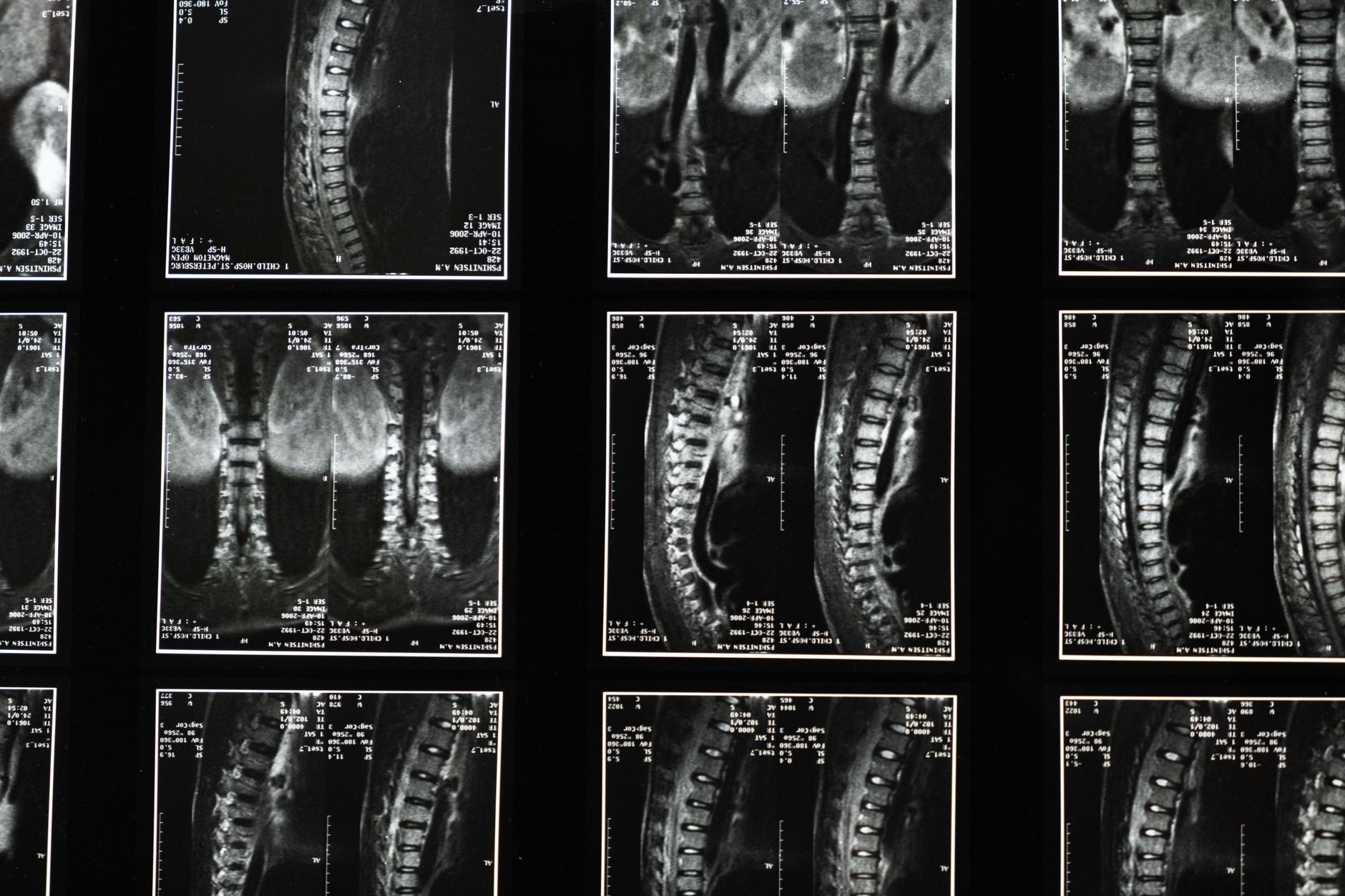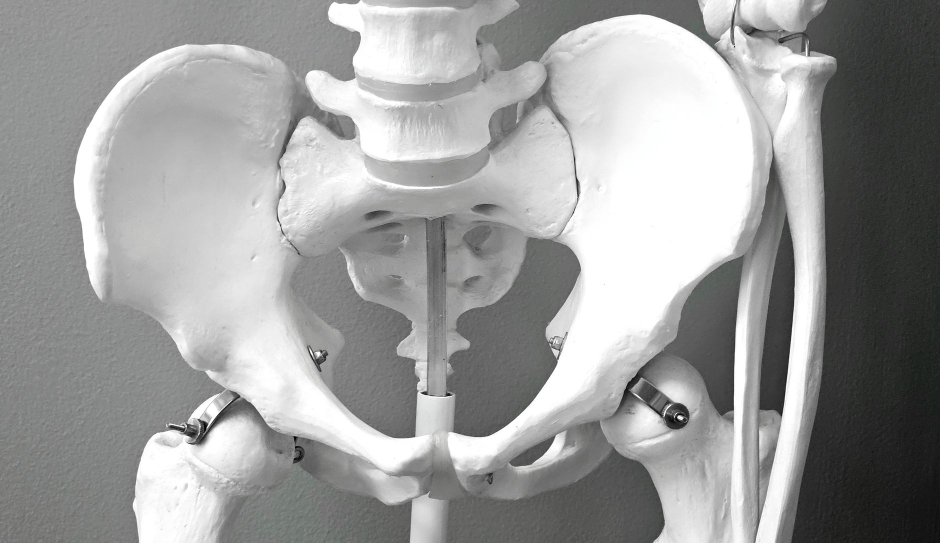

Lumbar spinal stenosis, a common cause of pain and disability in older adults, presents a persistent clinical paradox. Radiographic evidence of narrowing in the spinal canal often bears little resemblance to the severity of a patient's reported symptoms, or even whether they have symptoms at all. This disconnect complicates diagnosis, treatment planning, and patient expectations.
Spinal stenosis, particularly in the lumbar region, involves the narrowing of the spinal canal, nerve root canals, or intervertebral foramina. This narrowing can impinge on the spinal cord or nerve roots, theoretically leading to symptoms such as neurogenic claudication, radicular pain, and motor weakness. But the presence of these anatomical changes on imaging, whether X-ray, CT, or MRI, does not consistently correlate with the patient's clinical presentation.
Many individuals, particularly those over 50, exhibit significant degenerative changes on spinal imaging without experiencing any pain or functional limitations. Conversely, some patients with debilitating symptoms show only mild or equivocal findings on their scans. This discrepancy highlights the complexity of spinal pain and the limitations of relying solely on structural pathology.
The anatomical disconnect
The human spine undergoes continuous degenerative changes with age, a process that begins much earlier than most people realise. Disc desiccation, facet joint hypertrophy, ligamentum flavum thickening, and osteophyte formation are all common findings in asymptomatic adults. These changes collectively contribute to the narrowing of the spinal canal, yet they do not automatically translate into symptomatic compression.
One reason for this anatomical disconnect is the dynamic nature of the spinal canal. The degree of narrowing can vary with posture and activity. A patient might have significant stenosis in extension, which is often the position used during imaging, but experience less compression in flexion. This variability means a static image captures only a snapshot of a dynamic process, potentially overestimating or underestimating the functional impact of the narrowing.
Beyond structural compression
Pain perception in spinal stenosis is not solely a function of mechanical compression. Several other factors contribute to the symptom experience, including inflammation, vascular compromise, and central sensitisation. When nerve roots are compressed, local inflammatory mediators can be released, leading to pain even with relatively mild mechanical impingement. This inflammatory component may explain why some patients with minimal structural changes report severe pain.
Vascular compromise to the nerve roots, particularly during activity, also plays a role in neurogenic claudication. Even if the canal narrowing is not severe enough to cause direct mechanical damage, it can restrict blood flow to the nerve roots, leading to ischaemia and pain with exertion. This mechanism is often difficult to visualise directly on standard imaging, further decoupling the scan from the symptom.
The role of psychosocial factors
The experience of chronic pain, including that associated with spinal stenosis, is profoundly influenced by psychosocial factors. Depression, anxiety, fear avoidance behaviours, and catastrophising can all amplify pain perception and functional disability, irrespective of the underlying structural pathology. A patient's coping mechanisms and their perception of their condition can have a greater impact on their quality of life than the objective degree of canal narrowing.
Clinicians must consider these non-anatomical contributors when assessing patients. A detailed history and physical examination remain paramount, allowing for an understanding of the patient's pain experience, functional limitations, and psychological state. Relying solely on imaging risks misattributing symptoms to structural findings that may be incidental, leading to inappropriate or ineffective interventions. For a broader perspective on how imaging can reveal underlying conditions, consider how identifying rapid progressors in ADPKD often relies on specific imaging classifications.
Clinical implications for diagnosis and management
The poor correlation between imaging and symptoms means that a diagnosis of symptomatic spinal stenosis should always be primarily clinical. Imaging serves as a confirmatory tool, helping to identify the anatomical basis for symptoms that are consistent with the clinical picture. It should not be used as a screening tool in asymptomatic individuals, as this invariably leads to overdiagnosis and potentially unnecessary interventions.
When imaging does show significant stenosis in an asymptomatic patient, it is important to manage expectations and avoid alarm. These findings are often part of normal ageing. Similarly, if a patient presents with classic symptoms of neurogenic claudication but imaging shows only mild changes, the clinical picture should still guide treatment decisions. This approach aligns with general principles of musculoskeletal care, where a comprehensive assessment is prioritised, as detailed in resources like the Oxford Handbook of General Practice.
The challenge of incidental findings
Incidental findings on spinal imaging are exceedingly common. Studies consistently show that a large percentage of asymptomatic adults have disc bulges, herniations, and degenerative changes that would be considered pathological if found in a symptomatic patient. This prevalence of incidental findings makes it difficult to definitively link a specific imaging abnormality to a patient's pain, especially when symptoms are vague or non-specific.
The temptation to treat the image rather than the patient is a significant pitfall. Surgical interventions, for example, are most effective when there is a clear correlation between severe, functionally limiting symptoms and demonstrable structural compression. Performing surgery based solely on imaging findings in the absence of compelling symptoms often leads to suboptimal outcomes and patient dissatisfaction. The challenge of distinguishing clinical symptoms from subclinical inflammation is also a recurring theme in other fields, such as SpA remission.
Moving towards a holistic approach
Effective management of spinal stenosis requires a holistic approach that integrates clinical assessment, imaging findings, and psychosocial considerations. Non-surgical treatments, including physical therapy, exercise, pain medication, and epidural injections, are often the first line of therapy. These interventions aim to improve function, reduce pain, and enhance coping strategies, regardless of the precise degree of canal narrowing on a scan.
Patient education is also critical. Explaining the commonality of degenerative changes and the poor correlation between imaging and symptoms can help alleviate anxiety and set realistic expectations. It empowers patients to understand that their pain is real, even if the imaging does not provide a clear, singular explanation. This understanding is essential for managing chronic conditions, much like understanding the complex relationship of iron status and symptoms in polycythemia vera.
The goal is to improve the patient's quality of life and functional capacity, not simply to normalise an image. The limitations of imaging in spinal stenosis serve as a powerful reminder that the patient's lived experience, not just their anatomy, must remain at the centre of clinical decision-making. Future research will need to explore more dynamic imaging techniques or biomarkers that better capture the functional impact of spinal canal narrowing.
The persistent disconnect between imaging findings and patient symptoms in spinal stenosis should prompt a re-evaluation of diagnostic priorities. Relying heavily on MRI or CT scans for initial assessment risks over-medicalising age-related degenerative changes, potentially leading to unnecessary referrals and interventions for incidental findings.
Clinicians must instead sharpen their clinical acumen, prioritising a thorough history and physical examination to establish a clear clinical picture of neurogenic claudication or radicular pain. Imaging should serve as a confirmatory tool, used judiciously when symptoms align with suspected structural pathology, rather than a primary diagnostic filter.
This approach also necessitates a shift in patient communication. Explaining that degenerative changes are common and often asymptomatic can mitigate anxiety and manage expectations, preventing patients from fixating on a scan report that may not reflect their actual experience. It reinforces the idea that treatment targets function and pain relief, not merely a 'cleaner' image.
The field needs better tools to assess the functional impact of spinal narrowing, perhaps dynamic imaging or neurophysiological markers, to bridge this enduring gap between what we see on a screen and what a patient feels.
- The Pivot The anatomical changes visible on imaging do not reliably predict the presence or intensity of pain and functional impairment in spinal stenosis.
- The Data A significant proportion of asymptomatic individuals exhibit moderate to severe spinal canal narrowing on MRI.
- The Action Clinicians must prioritise a thorough clinical assessment over isolated imaging findings when evaluating and managing patients with suspected spinal stenosis.
ART-2026-1506
·10/26
Drafted with AI assistance, reviewed and approved by the editorial team. This publication is intended for healthcare professionals, researchers, and life science industry professionals. Content is provided for informational and educational purposes only and does not constitute medical advice.

Thirty years in health journalism, the last fifteen in life sciences. I have reported from every major medical congress and watched blockbuster drugs get revised after approval. I cover what the data says.
Cite This Article
Carter J, Voss M. Why spinal stenosis imaging often fails to explain patient symptoms. The Life Science Feed. Published August 27, 2026. Updated October 4, 2026. Accessed October 11, 2026. https://thelifesciencefeed.com/orthopedics/spinal-stenosis/insights/spinal-stenosis-imaging-symptoms-correlation.
Editorial & AI Standards
All content is researched from peer-reviewed, open-access sources: published trial data, clinical guidelines, and regulatory filings. AI tools are used solely to structure and summarise that evidence; no AI-generated conclusions appear without editor verification against the primary source.
Every article is reviewed by a named editor before publication. Source citations are listed in the References section. This content does not represent the views of any pharmaceutical company, medical device manufacturer, or healthcare provider.
Licence & Rights
© 2026 The Life Science Feed. All rights reserved. Unless otherwise indicated, all content is the property of The Life Science Feed and may not be reproduced, distributed, or transmitted in any form or by any means without prior written permission.
Medical Disclaimer
The information provided on The Life Science Feed is for educational and informational purposes only. It is not intended as a substitute for professional medical advice, diagnosis, or treatment. Always seek the advice of your physician or other qualified healthcare provider regarding any medical condition or treatment decision. Never disregard professional medical advice or delay in seeking it because of something you have read on this website.
References
1. Zhang L, Zeitoun D, Rangel A, Lazennec JY, Catonné Y, Pascal-Moussellard H. Preoperative evaluation of the cervical spondylotic myelopathy with flexion-extension magnetic resonance imaging: about a prospective study of fifty patients. Spine (Phila Pa 1976). 2011;36(17):E1134-9. doi:10.1097/BRS.0b013e3181f822c7