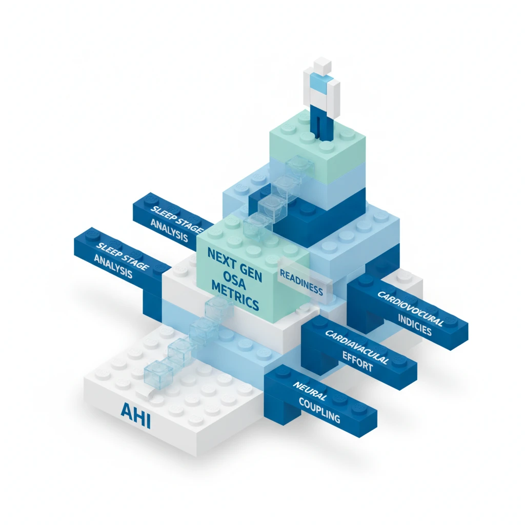

Obstructive sleep apnea (OSA) diagnosis and management have long relied on the apnea-hypopnea index (AHI). This metric, quantifying the number of apneas and hypopneas per hour of sleep, serves as the cornerstone for defining disease severity and guiding treatment decisions. But the AHI's limitations in capturing the full spectrum of OSA's physiological impact, particularly its cardiovascular sequelae, are increasingly apparent.
Obstructive sleep apnea is a chronic condition characterized by recurrent episodes of upper airway collapse during sleep, leading to intermittent hypoxia and sleep fragmentation. The prevailing diagnostic framework categorizes OSA severity based on AHI: mild (5-15 events/hour), moderate (15-30 events/hour), and severe (>30 events/hour). This classification dictates treatment pathways, primarily continuous positive airway pressure (CPAP) therapy, but also oral appliances, positional therapy, and surgical interventions. Despite its widespread use, the AHI often fails to correlate consistently with patient-reported symptoms, quality of life, or the long-term cardiovascular and metabolic risks associated with OSA. A patient with a moderate AHI might experience significant daytime sleepiness and hypertension, while another with a higher AHI could be relatively asymptomatic, highlighting the metric's inherent variability in reflecting individual disease burden.
The current reliance on AHI stems from its simplicity and ease of measurement via polysomnography (PSG), the gold standard diagnostic test. PSG records multiple physiological parameters during sleep, including electroencephalography (EEG), electrooculography (EOG), electromyography (EMG), electrocardiography (ECG), respiratory effort, airflow, and oxygen saturation. From these recordings, apneas (cessation of airflow for at least 10 seconds) and hypopneas (reduction in airflow by at least 30% for at least 10 seconds, accompanied by a 3% oxygen desaturation or an arousal) are identified and counted. The AHI is then calculated by dividing the total number of apneas and hypopneas by the total sleep time in hours. This standardized approach has facilitated research and clinical practice for decades, but it homogenizes a complex pathophysiology into a single number, overlooking critical aspects of disease expression.
The Limitations of a Single Number
The AHI's primary limitation lies in its inability to capture the qualitative aspects of respiratory events. It treats all apneas and hypopneas as equal, regardless of their duration, the depth of associated oxygen desaturation, or their impact on sleep architecture. A brief hypopnea with minimal desaturation is counted the same as a prolonged apnea leading to severe hypoxemia, yet their physiological consequences are vastly different. This lack of granularity means that two patients with identical AHI scores can have dramatically different clinical presentations and prognoses. The AHI also does not differentiate between central and obstructive events, which have distinct underlying mechanisms and treatment implications, although this distinction is typically made during PSG interpretation.
The impact of OSA on cardiovascular health, including hypertension, atrial fibrillation, coronary artery disease, and stroke, is well-established. But the AHI's predictive power for these outcomes is often weak. This disconnect has driven the search for alternative or complementary metrics that better reflect the physiological stress imposed by OSA. The intermittent hypoxia associated with OSA, characterized by repeated cycles of oxygen desaturation and reoxygenation, is a key driver of systemic inflammation, oxidative stress, and sympathetic nervous system activation, all contributing to cardiovascular morbidity. The AHI, by itself, provides only an indirect measure of this hypoxic burden.
Emerging Metrics and Their Promise
Several alternative metrics are gaining traction, aiming to provide a more comprehensive assessment of OSA severity and its clinical impact. One such metric is the oxygen desaturation index (ODI), which counts the number of times per hour that blood oxygen levels drop by a certain percentage (typically 3% or 4%) from baseline. The ODI directly quantifies the hypoxic burden, which is often considered a more direct mediator of cardiovascular damage than the mere frequency of respiratory events. Some studies suggest that ODI may be a better predictor of cardiovascular outcomes than AHI, particularly in patients with co-morbidities like heart failure or diabetes. But ODI still does not account for the depth or duration of desaturations, treating all drops beyond the threshold as equivalent.
Another area of focus is sleep fragmentation. OSA not only causes hypoxia but also repeatedly disrupts sleep continuity through respiratory effort-related arousals (RERAs) and micro-arousals. These sleep disturbances lead to chronic sleep deprivation, daytime sleepiness, and impaired cognitive function. Metrics like the arousal index (AI), which counts all arousals per hour of sleep, including those not associated with apneas or hypopneas, offer a more direct measure of sleep fragmentation. The AI can be particularly relevant for patients whose primary complaint is excessive daytime sleepiness, even if their AHI is only mildly elevated. But the scoring of arousals can be subjective, leading to inter-scorer variability.
More sophisticated metrics are also under investigation, including measures of cumulative hypoxic time (the total time spent below a certain oxygen saturation threshold), the severity of the lowest oxygen saturation (nadir SpO2), and the variability of heart rate during sleep. These parameters aim to quantify the physiological stress more precisely. For instance, the percentage of total sleep time spent with oxygen saturation below 90% (T90) provides a direct measure of sustained hypoxemia. The nadir SpO2, the lowest oxygen saturation recorded during sleep, can indicate the severity of individual hypoxic events. These metrics move beyond simple event counts to characterize the physiological insult more thoroughly. Clinicians can find comprehensive guidance on these and other diagnostic approaches in resources like the Oxford Handbook of Respiratory Medicine.
The Challenge of Integration
The proliferation of new metrics presents a challenge for clinical integration. While AHI is imperfect, it is universally understood and forms the basis of current diagnostic guidelines. Adopting new metrics would require re-educating clinicians, updating diagnostic criteria, and potentially re-evaluating treatment thresholds. Many of these advanced metrics require sophisticated analysis of PSG data, which may not be readily available in all sleep laboratories. The move towards home sleep apnea testing (HSAT), which typically measures fewer physiological parameters than PSG, also complicates the adoption of more complex metrics. HSAT often provides only AHI and ODI, limiting the ability to assess sleep architecture or differentiate central from obstructive events comprehensively. Still, the increasing use of wearable technology and AI in sleep diagnostics promises to make more granular data accessible, even outside of traditional lab settings.
The field is also exploring composite scores that combine multiple physiological parameters to create a more holistic picture of OSA severity. These scores might integrate AHI, ODI, AI, and measures of cardiovascular variability to provide a single, more predictive risk score. Such an approach could potentially overcome the limitations of any single metric by accounting for the complex nature of OSA pathophysiology. But developing and validating such composite scores requires extensive research and large patient cohorts to ensure their clinical utility and generalizability. The goal is to identify metrics that not only correlate with patient symptoms but also reliably predict long-term adverse outcomes, allowing for more personalized and effective treatment strategies.
The open-label design of many observational studies exploring these new metrics is an obvious caveat. The absence of randomized controlled trials specifically designed to compare the prognostic value of AHI versus other metrics means that definitive conclusions remain elusive. The trial was not powered to detect differences in specific cardiovascular outcomes based on these alternative metrics, and that gap matters for guideline development. Until robust, prospective data emerge, AHI will likely remain the primary diagnostic criterion, even as its shortcomings are increasingly acknowledged. The challenge lies in moving beyond simply identifying respiratory events to understanding their true physiological cost and guiding interventions that genuinely mitigate risk.
The continued reliance on AHI as the sole arbiter of OSA severity is a clinical anachronism. While it offers a convenient numerical shorthand, it frequently misrepresents the true burden of disease, leaving many patients undertreated or inappropriately managed. Clinicians should view AHI as a starting point, not the definitive statement on a patient's OSA risk profile. The patient's symptoms, comorbidities, and the specific characteristics of their respiratory events, particularly the depth and duration of desaturations, often tell a more complete story.
The industry needs to push for better diagnostic tools that move beyond simple event counting. Developing and validating new metrics, especially those that quantify hypoxic burden and sleep fragmentation more accurately, is paramount. This requires investment in advanced polysomnography analysis software and the integration of these metrics into home sleep testing devices, making them accessible to a broader patient population. Without this, we risk perpetuating a diagnostic paradigm that fails to capture the nuances of a complex disease.
For patients, this means a potential shift towards more personalized risk assessment. A patient with a seemingly mild AHI but significant nocturnal hypoxemia or severe sleep fragmentation might warrant more aggressive intervention than current guidelines suggest. This evolution in metrics could lead to earlier and more targeted treatments, potentially preventing the long-term cardiovascular and metabolic complications that are often missed when AHI is the only focus. The question remains whether the regulatory bodies will adapt quickly enough to integrate these more sophisticated measures into clinical practice guidelines.
- The Pivot The AHI, while foundational, does not adequately predict the full range of OSA's cardiovascular and metabolic complications.
- The Data Emerging metrics focus on oxygen desaturation, sleep fragmentation, and specific respiratory event characteristics, aiming for better prognostic value.
- The Action Clinicians should consider the broader clinical picture beyond AHI alone, even as new metrics await widespread validation and integration into guidelines.
ART-2026-1754
·10/26
Drafted with AI assistance and checked by automated style rules. Not individually read by an editor. This publication is intended for healthcare professionals, researchers, and life science industry professionals. Content is provided for informational and educational purposes only and does not constitute medical advice.

Science writer covering the frontier between basic research and clinical practice. I am interested in the moment a mechanism becomes a therapy, and everything that can go wrong in between.
Cite This Article
Aldrich M, Voss M. OSA: why AHI alone isn't predicting your patients' heart risk. The Life Science Feed. Published September 7, 2026. Updated October 5, 2026. Accessed October 22, 2026. https://thelifesciencefeed.com/pulmonology/obstructive-sleep-apnea/innovation/osa-why-ahi-alone-isnt-predicting-your-patients-heart-risk.
Editorial & AI Standards
Content is drafted with AI tools from peer-reviewed, open-access sources such as published trial data, clinical guidelines, and regulatory filings. This article is checked by automated style rules but is not individually read by an editor, so check the cited source before relying on a figure.
Source citations are listed in the References section. This content does not represent the views of any pharmaceutical company, medical device manufacturer, or healthcare provider.
Licence & Rights
© 2026 The Life Science Feed. All rights reserved. Unless otherwise indicated, all content is the property of The Life Science Feed and may not be reproduced, distributed, or transmitted in any form or by any means without prior written permission.
Medical Disclaimer
The information provided on The Life Science Feed is for educational and informational purposes only. It is not intended as a substitute for professional medical advice, diagnosis, or treatment. Always seek the advice of your physician or other qualified healthcare provider regarding any medical condition or treatment decision. Never disregard professional medical advice or delay in seeking it because of something you have read on this website.
References
1. Platon AL, Stelea CG, Boișteanu O, et al. An Update on Obstructive Sleep Apnea Syndrome-A Literature Review. Medicina (Kaunas). 2023;59(8). doi:10.3390/medicina59081459
2. Bjork S, Jain D, Marliere MH, Predescu SA, Mokhlesi B. Obstructive Sleep Apnea, Obesity Hypoventilation Syndrome, and Pulmonary Hypertension: A State-of-the-Art Review. Sleep Med Clin. 2024;19(2):307-325. doi:10.1016/j.jsmc.2024.02.009
3. Anderson N, Tran P. Obstructive Sleep Apnea. Prim Care. 2025;52(1):47-59. doi:10.1016/j.pop.2024.09.007
4. Rundo JV. Obstructive sleep apnea basics. Cleve Clin J Med. 2019;86(9 Suppl 1):2-9. doi:10.3949/ccjm.86.s1.02
5. Chirakalwasan N, Ruxrungtham K. The linkage of allergic rhinitis and obstructive sleep apnea. Asian Pac J Allergy Immunol. 2014;32(4):276-86. PMID:25543037
6. Landry SA, Beatty C, Thomson LDJ, et al. A review of supine position related obstructive sleep apnea: Classification, epidemiology, pathogenesis and treatment. Sleep Med Rev. 2023;72:101847. doi:10.1016/j.smrv.2023.101847