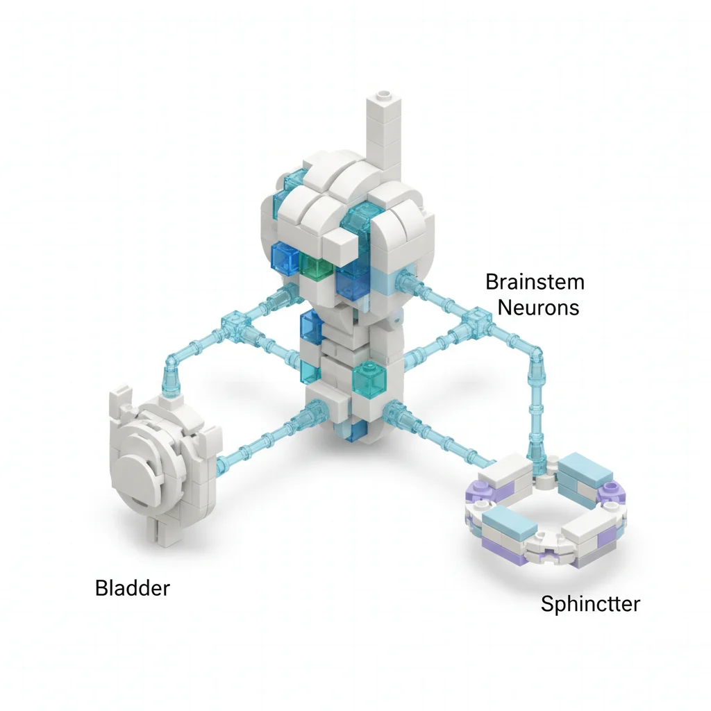

Urination requires a finely tuned dance between bladder contraction and external urethral sphincter (EUS) relaxation. When this coordination fails, as in detrusor-sphincter dyssynergia, patients face significant morbidity. For years, the specific neuronal subpopulations orchestrating this vital process remained elusive, hindering targeted therapeutic development. A new study published in Elife identifies a key cell type in the brainstem that controls this bladder-urethra coordination.1
The mammalian urinary system relies on a complex neural network to manage urine storage and expulsion. This network ensures that the bladder contracts while the external urethral sphincter relaxes, a synchronized event essential for efficient voiding. Dysregulation of this intricate coordination leads to conditions like detrusor-sphincter dyssynergia, a common and debilitating issue in patients with neurological disorders such as spinal cord injury or multiple sclerosis.1,3 Identifying the precise neuronal populations responsible for this coordination has been a long-standing challenge in neuro-urology, limiting the development of specific, effective treatments.1,2
Investigators, including Li X, Li X, and Li J, set out to pinpoint the specific subpopulation of neurons within the pontine micturition center (PMC) that orchestrates this bladder-EUS synergy. The PMC, located in the brainstem, has long been recognized as a critical relay station for micturition reflexes. But the exact cellular players within this region remained poorly defined. The research, published in 2026, utilized advanced genetic and physiological techniques in animal models to isolate and characterize these neurons.1
Pinpointing the PMC's Conductors
The study focused on cells expressing estrogen receptor 1 (ESR1+) within the PMC. These PMCESR1+ neurons emerged as central figures in the voiding reflex. When these specific neurons were activated, voiding initiated with 100% reliability. Conversely, when the PMCESR1+ neurons were suppressed, ongoing voiding was suspended with 100% reliability. This level of control suggests a direct and indispensable role for these cells in the micturition process.1
The researchers further dissected the neural pathways involved. They performed selective nerve transections to understand how PMCESR1+ neurons communicate with the bladder and the EUS. Transection of the pelvic nerve, which primarily innervates the bladder, did not impair the PMCESR1+ neurons' control over the EUS via the pudendal nerve. This indicates a distinct and independent pathway for sphincter control. Conversely, transection of the pudendal nerve, which controls the EUS, did not affect the PMCESR1+ neurons' control over the bladder via the pelvic nerve. This anatomical separation of control pathways, while originating from a common brainstem center, highlights the sophisticated parallel processing involved in urination.1
Anatomy of Coordination
The anatomical investigation revealed that PMCESR1+ neurons are not a monolithic population. They consist of three distinct subpopulations, categorized by their spinal projection patterns. One subpopulation targets the sacral parasympathetic nucleus, which is essential for bladder contraction. Another innervates the dorsal gray commissure, a region implicated in somatic motor control, including the EUS. A third subpopulation projects to both regions, effectively acting as an integrative hub. This tripartite organization enforces the coordination of bladder contraction and sphincter relaxation in a rigid temporal sequence, ensuring efficient and complete voiding.1
This detailed neuroanatomical mapping provides a cellular-level understanding of how the brainstem coordinates these seemingly disparate functions. The existence of a subpopulation projecting to both bladder and sphincter control centers suggests a hardwired mechanism for synchronicity, preventing the dyssynergia seen in various neurological conditions. The maturation of these micturition-related neural circuits in postnatal rats, as explored by Nitabara A and colleagues in 2025, further reinforces the developmental importance of these pathways.2 The Oxford Handbook of Neurology offers a concise overview of such complex neural circuits and their clinical relevance.
Implications for Dyssynergia
Detrusor-sphincter dyssynergia (DSD) is a condition where the bladder contracts against a closed or inadequately relaxed EUS, leading to incomplete voiding, high bladder pressures, and potential kidney damage. Current treatments often involve broad neuromodulation or symptomatic management, which can have off-target effects or limited efficacy. The identification of PMCESR1+ neurons as the precise coordinators offers a potential target for highly specific interventions.1,3
For instance, therapies could be developed to modulate the activity of these specific ESR1+ neurons, either by enhancing their function in cases of underactive bladder or by fine-tuning their coordination in DSD. This precision contrasts sharply with current approaches that often involve botulinum toxin injections into the detrusor or EUS, or sacral neuromodulation, which lack the cellular specificity now identified. The detailed neuroanatomy of the pontine micturition center, as reviewed by Rahman M and Siddik AB in 2026, reinforces the complexity and the need for such targeted approaches.3
Limitations and Future Directions
The primary limitation of this research is its reliance on animal models. While the micturition reflex is conserved across mammals, direct translation to human physiology always carries caveats. The exact molecular and cellular characteristics of human PMCESR1+ neurons and their precise connectivity may differ. Further studies are needed to confirm these findings in human tissues and to develop methods for non-invasive or minimally invasive modulation of these specific neuronal populations in a clinical setting.1
The study also did not explore the upstream inputs that regulate PMCESR1+ neuron activity. Understanding how higher brain centers, such as the prefrontal cortex or insula, influence these brainstem neurons could provide additional targets for treating voiding dysfunction, particularly in conditions with a strong psychological component or voluntary control issues. The role of estrogen signaling itself in these neurons also warrants further investigation, given the expression of ESR1. This could have implications for sex-specific differences in voiding dysfunction or the impact of hormonal therapies.1
The next logical step involves developing tools to specifically target PMCESR1+ neurons in preclinical models of detrusor-sphincter dyssynergia. This could involve viral vectors for gene therapy or novel pharmacological agents that selectively act on these cells. Proving that modulation of these neurons can reverse DSD symptoms in animal models would be a critical bridge to human clinical trials. The long-term safety and efficacy of such highly specific interventions would then need rigorous evaluation.1
The identification of PMCESR1+ neurons as the precise conductors of bladder-sphincter coordination is a significant step forward for neuro-urology. For clinicians managing patients with detrusor-sphincter dyssynergia, this research offers a much-needed shift from symptomatic relief to potential disease modification. Current treatments often involve a trial-and-error approach with limited success, leaving many patients with chronic catheterization or significant quality of life impairment.
This work provides a clear, anatomically defined target. Instead of broadly modulating sacral nerves or injecting botulinum toxin with its systemic absorption risks, future therapies could aim to activate or suppress these specific brainstem neurons. This precision could minimize off-target effects and improve therapeutic ratios, a welcome development for a patient population often burdened by polypharmacy and complex neurological comorbidities.
The role of estrogen receptor 1 expression also raises interesting questions. While the study did not examine the hormonal regulation of these neurons, it suggests a potential link between endocrine status and voiding function. This might explain some observed sex differences in bladder control disorders and could open avenues for hormonal modulators, though much more research is needed before any clinical recommendations can be made.
Still, the path from animal model to human therapy is long and fraught with challenges. The complexity of the human brainstem and the ethical considerations of direct brainstem interventions mean that clinical translation will require innovative delivery methods and extensive safety validation. But for now, this research provides a solid neurobiological foundation upon which truly targeted therapies for urination disorders can begin to be built.
- The Pivot Specific ESR1+ neurons in the pontine micturition center (PMC) are the precise coordinators of bladder-EUS function.
- The Data Activation of these PMCESR1+ neurons initiates voiding with 100% reliability; suppression suspends it with 100% reliability.1
- The Action Understanding this specific neuronal population opens avenues for highly targeted therapies for urination disorders, moving beyond broad neuromodulation.
ART-2026-1098
·08/26
Drafted with AI assistance, reviewed and approved by the editorial team. This publication is intended for healthcare professionals, researchers, and life science industry professionals. Content is provided for informational and educational purposes only and does not constitute medical advice.

I cover NHS policy, NICE guidance, and the gap between what the evidence says and what gets commissioned. I bring a health economics background to reporting on how health systems make decisions under uncertainty.
Cite This Article
Mistry D, Lopes W. Bladder-sphincter dyssynergia: a brainstem switch just flipped our understanding. The Life Science Feed. Published August 13, 2026. Updated August 13, 2026. Accessed August 13, 2026. https://thelifesciencefeed.com/urology/urinary-incontinence/research/bladder-sphincter-dyssynergia-a-brainstem-switch-just-flipped-our-understanding.
Editorial & AI Standards
All content is researched from peer-reviewed, open-access sources: published trial data, clinical guidelines, and regulatory filings. AI tools are used solely to structure and summarise that evidence; no AI-generated conclusions appear without editor verification against the primary source.
Every article is reviewed by a named editor before publication. Source citations are listed in the References section. This content does not represent the views of any pharmaceutical company, medical device manufacturer, or healthcare provider.
Licence & Rights
© 2026 The Life Science Feed. All rights reserved. Unless otherwise indicated, all content is the property of The Life Science Feed and may not be reproduced, distributed, or transmitted in any form or by any means without prior written permission.
Medical Disclaimer
The information provided on The Life Science Feed is for educational and informational purposes only. It is not intended as a substitute for professional medical advice, diagnosis, or treatment. Always seek the advice of your physician or other qualified healthcare provider regarding any medical condition or treatment decision. Never disregard professional medical advice or delay in seeking it because of something you have read on this website.
References
1. Li X, Li X, Li J. Brainstem neurons coordinate the bladder and urethral sphincter for urination. Elife. 2026;15:e42559936. https://pubmed.ncbi.nlm.nih.gov/42559936/
2. Nitabara A, Banjo Y, Takiguchi M. Maturation of Micturition-Related Neural Circuits That Control Pelvic Visceromotor Functions in Postnatal Rats. Eur J Neurosci. 2025;61(1):20-35. https://pubmed.ncbi.nlm.nih.gov/40922463/
3. Rahman M, Siddik AB. Neuroanatomy, Pontine Micturition Center. In: StatPearls. StatPearls Publishing; 2026. https://pubmed.ncbi.nlm.nih.gov/32491351/